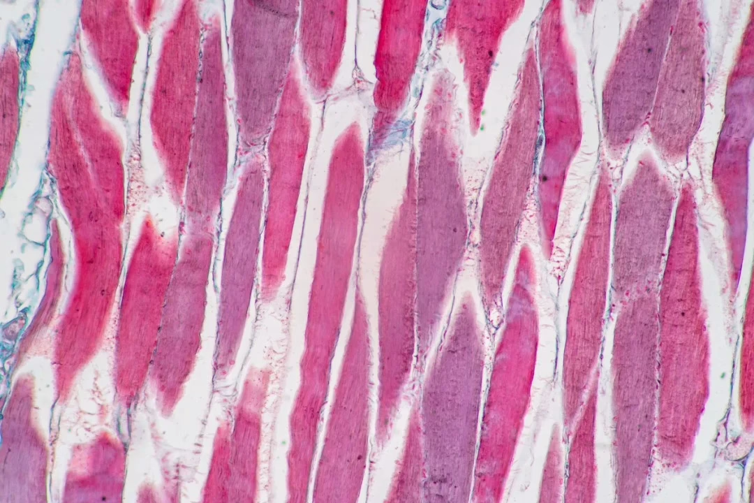
This compound has garnered attention from researchers for its hypothesized potential to interfere with myostatin signaling. Myostatin signaling is a critical pathway that regulates the development of skeletal muscle cells. The observable interactions between Ramatercept and myostatin when both are exposed to laboratory models are believed to disrupt downstream signaling events. These downstream signaling events are believed to generally limit muscular tissue growth by inhibiting the proliferation of satellite cells and protein synthesis. Preclinical studies conducted by researchers exposing these proteins to genetically modified murine models have produced data suggesting that inactivating ActRIIB may lead to a substantial increase in skeletal muscle cell volume. This may encourage further investigation into the physiological roles of Ramatercept.
Beyond skeletal muscle cell biology, activin signaling is believed to be involved in various cellular processes. This is thought to include general reproductive development and oncogenesis. Alterations in ActRIIB activity have been documented in certain malignancies, such as colorectal and prostate cancers, as well as in murine testicular tissue. In these instances, the receptor may play a role in regulating spermatogenesis. Some data suggests a hypothetical modulatory impact of ActRIIB ligands on adipose tissue deposition and bone mineral homeostasis. That said, the molecular mechanisms underlying these impacts have not yet been fully explored.
Contents:
- Mechanisms of Action
- Scientific and Research Studies
- Ramatercept (ACE-031) and Skeletal Muscle Function
- Ramatercept (ACE-031) and Tissue Preservation
- Ramatercept (ACE-031) and Muscle Cell Energy Dynamics
- Ramatercept (ACE-031) and Duchenne Muscular Dystrophy
- Ramatercept (ACE-031) and Bone Physiology
- Ramatercept (ACE-031) in Cancer-Associated Muscular Tissue Wasting
- References
Mechanism of Action
The functional model of Ramatercept centers on its interaction with multiple inhibitory ligands that act upstream of muscle cell signaling. Research suggests that Ramatercept binds not only to myostatin but also to other members of the TGF-β superfamily, including activin A, BMP-2, and BMP-7. These proteins are considered to collectively contribute to the suppression of skeletal muscle growth by activating SMAD2/3-dependent transcriptional programs that negatively regulate anabolic processes.
In comparative studies of murine models, introduction of a soluble ActRIIB-Fc construct led to greater increases in muscle mass than agents designed to neutralize myostatin alone. This impact was observed even in genetically engineered murine models that lack functional myostatin, suggesting that other ligands may play parallel or compensatory roles in restricting muscle cell hypertrophy. The implication is that Ramatercept may exert its impacts by broadly neutralizing multiple antagonistic signals, thereby shifting the balance toward muscle cell anabolism.
This ligand-trapping strategy may also interrupt feedback loops within the TGF-β network, which are thought to fine-tune responses to metabolic and inflammatory cues. By disrupting these inhibitory pathways at the receptor level, Ramatercept introduces a potential tool for exploring multi-pathway regulation of muscular tissue mass in degenerative and catabolic contexts.[1]
Scientific Research and Studies
Ramatercept (ACE-031) and Skeletal Muscle Function
Preclinical investigations in murine models suggest that exposure of these models to Ramatercept may support the contractile performance of skeletal muscle. Observed increases in both peak and cumulative force output, approximately 40% and 25% respectively, suggest that the compound may impact the intrinsic mechanical properties of muscular tissue fibers.[1] These impacts appear to occur without significant changes in muscle cell fatigue parameters, implying that functional gains may be dissociated from alterations in energy depletion kinetics.
Further metabolic analyses revealed that neither ATP availability nor overall contractile efficiency underwent measurable changes following Ramatercept exposure. This finding suggests that the support in force generation is unlikely to stem from modifications in energy metabolism, but rather from changes in excitation-contraction coupling or architecture of muscular tissue fibers. Data from these models suggest a potential role for Ramatercept in selectively supporting existing mechanical strength, while also highlighting its ability to maintain energetic stability within muscular tissue.[2]
Ramatercept (ACE-031) and Tissue Preservation
A controlled clinical study involving postmenopausal female participants examined the impacts of Ramatercept introduction on musculoskeletal composition.[3]
The findings suggest a statistically significant increase in lean muscular tissue mass and volume within 29 days post-intervention, indicating that the agent may exert protective or anabolic impacts in otherwise functional muscular tissue.
In addition to musculoskeletal outcomes, the study identified notable shifts in biochemical markers associated with bone formation and adipose metabolism. These secondary impacts imply broader physiological involvement of Ramatercept beyond its interaction with muscle cell-related pathways. Investigators reported a potential downregulation of fat deposition processes in parallel with support of bone matrix turnover, as reflected in circulating biomarker profiles.
Collectively, these results support the hypothesis that Ramatercept may act through a constellation of pathways involving not only skeletal muscle but also bone and adipose tissue regulation. While mechanistic details remain under investigation, the emerging data positions Ramatercept as a candidate for broader study in systemic tissue remodeling contexts.
Ramatercept (ACE-031) and Muscle Cell Energy Dynamics
Experimental data from murine models suggest that myostatin acts as a negative modulator of muscle cell energy homeostasis. Elevated myostatin expression has been associated with mitigated endurance and increased susceptibility to metabolic fatigue. In models where endogenous myostatin was disrupted, specifically via activin type IIB receptor (ActRIIB) blockade, significant elevations in serum lactate concentrations and histological data of metabolic injury to muscular tissue fibers were observed.
Interestingly, this intervention also appeared to diminish skeletal muscle capillarization, potentially mitigating the efficiency of oxygen delivery and contributing to the observed energetic stress. The same laboratory conditions indicated potential downregulation of molecular markers central to mitochondrial oxidative function, including Pparβ, Pgc1α, and Pdk4. These alterations suggest that ActRIIB signaling plays a regulatory role in preserving oxidative metabolism and vascular integrity in muscular tissue.[4]
Conversely, Ramatercept, a soluble ActRIIB-Fc fusion protein, may mitigate these impacts by sequestering extracellular myostatin and related ligands. This mechanism may facilitate an increase in oxidative metabolic capacity, thereby mitigating the accumulation of fatigue-inducing byproducts during muscle cell activity. A better-supported oxidative potential in skeletal muscle may also mitigate the detrimental impacts of reactive oxygen species generated under stress conditions.
Ramatercept (ACE-031) and Duchenne Muscular Dystrophy
Duchenne muscular dystrophy (DMD) is an X-linked recessive neuromuscular disorder marked by progressive and severe muscle degeneration. Impacted organisms often exhibit loss of ambulation by early adolescence. At the histological level, skeletal muscle fibers in DMD indicate significantly diminished contractile protein content and an abnormal accumulation of intramuscular fat. The primary etiological factor is the absence or dysfunction of dystrophin, a cytoskeletal protein that stabilizes the muscle fiber membrane during contraction cycles.
In addition to the mechanical instability conferred by dystrophin deficiency, secondary molecular events may contribute to disease progression. Among these is the increased release of myostatin, a member of the transforming growth factor-β (TGF-β) superfamily, from damaged myofibers. Elevated local myostatin concentrations may suppress regenerative signaling in adjacent muscle progenitor cells, further impairing recovery and contributing to muscle wasting.
Although gene-based strategies for dystrophin replacement continue to encounter translational barriers, modulation of myostatin signaling has emerged as an adjunctive agent for study. Ramatercept, a recombinant fusion protein based on the extracellular domain of activin type IIB receptor (ActRIIB), has been evaluated for its potential to interfere with myostatin-mediated signaling and support muscle cell maintenance in DMD models.
A clinical study reported by Campbell et al.[5] explored the impacts of Ramatercept on DMD-related muscle cell decline. In this trial, the findings suggested that the introduction of the agent was associated with the preservation of motor performance, including stabilization of six-minute walk distances. Researchers also observed increases in lean tissue mass, support for bone mineral density, and mitigation in fat mass. These secondary outcomes are consistent with prior findings suggesting that ActRIIB inhibition has broader systemic impacts on musculoskeletal and adipose tissue regulation.
The study concluded that inhibition of the myostatin signaling axis with Ramatercept may offer a mechanistically plausible pathway for further study, in an effort to counteract both primary and secondary muscle cell pathology.
Ramatercept (ACE-031) and Bone Physiology
Preclinical investigations using murine models of Duchenne muscular dystrophy (DMD) have provided data suggesting that Ramatercept may exert ancillary impacts on skeletal function.
In one study,[6] weekly peptide exposure over seven weeks resulted in notable increases in total muscular tissue mass, skeletal muscle weight, and bone mineral density. The support in bone parameters appeared to coincide with a mitigation in osteoclast population, cells primarily involved in bone resorption. Additionally, biomechanical analysis revealed support for the structural properties of the bone, such as an increased maximum force threshold and stiffness, suggesting potential alterations in bone microarchitecture.
While the initial focus of these investigations centered on muscular hypertrophy, the observed bone-specific outcomes have prompted interest in the broader skeletal implications of Ramatercept. The peptide’s apparent impact on osteoclastogenesis suggests a mechanistic divergence from myostatin inhibition alone, possibly involving the modulation of additional signaling pathways.
Further studies have examined this hypothesis more directly. In a separate murine model study,[7] researchers compared the impacts of Ramatercept with those of a myostatin-specific antagonist and a placebo control. Although both experimental agents were associated with supporting muscular tissue mass, only Ramatercept was associated with a marked increase in bone mineral density. This included a reported 132% increase in femoral density and a 27% increase in vertebral bone density, an observation that suggests Ramatercept may modulate bone turnover via myostatin-independent mechanisms.
These findings suggest that Ramatercept may interact with a broader range of ligands within the TGF-β superfamily, resulting in multifaceted impacts on musculoskeletal integrity.
Ramatercept (ACE-031) in Cancer-Associated Muscular Tissue Wasting
In vitro studies have also suggested that Ramatercept may play a role in modulating energy metabolism and preserving skeletal muscle integrity under oncologic stress. Specifically, data suggest that Ramatercept might attenuate activation of the ERK1/2 signaling cascade in cultured myotubes, a pathway implicated in apoptotic muscle cell death. By mitigating signaling through this axis, Ramatercept may delay or mitigate myofiber atrophy under catabolic conditions.
Mitochondrial preservation also appears to be a contributing mechanism. Research suggests that Ramatercept supports oxidative energy efficiency in muscle cells, which may be relevant in the metabolic context of cancer. Malignant cells often exert paracrine impacts that impair muscle cell mitochondrial function, mitigate ATP synthesis, and promote the production of reactive oxygen species (ROS). By supporting mitochondrial integrity, Ramatercept may mitigate these impacts and support cellular energy balance even under nutrient-deficient states.[8]
These findings have broader implications. Inhibition of myostatin via Ramatercept may contribute to the maintenance of lean muscular tissue mass and overall function in cachectic states. Moreover, some reports suggest that modulating this pathway may also impact systemic metabolic processes, including supporting insulin sensitivity, limiting ectopic fat accumulation, and supporting bone remodeling and fracture repair. Given the multifactorial points of potential relevance suggested by preclinical data, some researchers have advocated further exploration of Ramatercept as a supportive agent in combination with cytotoxic studies, particularly in mitigating chemotherapy-induced sarcopenia.
Disclaimer: The products mentioned are not intended for human or animal consumption. Research chemicals are intended solely for laboratory experimentation and/or in-vitro testing. Bodily introduction of any sort is strictly prohibited by law. All purchases are limited to licensed researchers and/or qualified professionals. All information shared in this article is for educational purposes only.
References:
- Cadena SM, Tomkinson KN, Monnell TE, Spaits MS, Kumar R, Underwood KW, Pearsall RS, Lachey JL. Administration of a soluble activin type IIB receptor promotes skeletal muscle growth independent of fiber type. J Appl Physiol (1985). 2010 Sep;109(3):635-42. doi: 10.1152/japplphysiol.00866.2009. Epub 2010 May 13. PMID: 20466801; PMCID: PMC2944638. https://www.ncbi.nlm.nih.gov/pmc/articles/PMC2944638/
- Béchir N, Pecchi E, Vilmen C, Le Fur Y, Amthor H, Bernard M, Bendahan D, Giannesini B. ActRIIB blockade increases force-generating capacity and preserves energy supply in exercising mdx mouse muscle in vivo. FASEB J. 2016 Oct;30(10):3551-3562. doi: 10.1096/fj.201600271RR. Epub 2016 Jul 14. PMID: 27416839. https://pubmed.ncbi.nlm.nih.gov/27416839/
- Attie KM, Borgstein NG, Yang Y, Condon CH, Wilson DM, Pearsall AE, Kumar R, Willins DA, Seehra JS, Sherman ML. A single ascending-dose study of muscle regulator Ramatercept (ACE-031) in healthy volunteers. Muscle Nerve. 2013 Mar;47(3):416-23. doi: 10.1002/mus.23539. Epub 2012 Nov 21. PMID: 23169607. https://pubmed.ncbi.nlm.nih.gov/23169607/
- Relizani K, Mouisel E, Giannesini B, Hourdé C, Patel K, Morales Gonzalez S, Jülich K, Vignaud A, Piétri-Rouxel F, Fortin D, Garcia L, Blot S, Ritvos O, Bendahan D, Ferry A, Ventura-Clapier R, Schuelke M, Amthor H. Blockade of ActRIIB signaling triggers muscle fatigability and metabolic myopathy. Mol Ther. 2014 Aug;22(8):1423-1433. doi: 10.1038/mt.2014.90. Epub 2014 May 27. PMID: 24861054; PMCID: PMC4435590. https://pubmed.ncbi.nlm.nih.gov/24861054/
- Campbell C, McMillan HJ, Mah JK, Tarnopolsky M, Selby K, McClure T, Wilson DM, Sherman ML, Escolar D, Attie KM. Myostatin inhibitor Ramatercept (ACE-031) treatment of ambulatory boys with Duchenne muscular dystrophy: Results of a randomized, placebo-controlled clinical trial. Muscle Nerve. 2017 Apr;55(4):458-464. doi: 10.1002/mus.25268. Epub 2016 Dec 23. PMID: 27462804. https://pubmed.ncbi.nlm.nih.gov/27462804/
- Puolakkainen T, Ma H, Kainulainen H, Pasternack A, Rantalainen T, Ritvos O, Heikinheimo K, Hulmi JJ, Kiviranta R. Treatment with soluble activin type IIB-receptor supports bone mass and strength in a mouse model of Duchenne muscular dystrophy. BMC Musculoskelet Disord. 2017 Jan 19;18(1):20. doi: 10.1186/s12891-016-1366-3. PMID: 28103859; PMCID: PMC5244551. https://www.ncbi.nlm.nih.gov/pmc/articles/PMC5244551/
- Bialek P, Parkington J, Li X, Gavin D, Wallace C, Zhang J, Root A, Yan G, Warner L, Seeherman HJ, Yaworsky PJ. A myostatin and activin decoy receptor supports bone formation in mice. Bone. 2014 Mar;60:162-71. doi: 10.1016/j.bone.2013.12.002. Epub 2013 Dec 9. PMID: 24333131. https://pubmed.ncbi.nlm.nih.gov/24333131/
- Lokireddy S, Wijesoma IW, Bonala S, Wei M, Sze SK, McFarlane C, Kambadur R, Sharma M. 1) Myostatin is a novel tumoral factor that induces cancer cachexia. Biochem J. 2015 Feb 15;466(1):201. doi: 10.1042/bj4660201u. Erratum for: Biochem J. 2012 Aug 15;446(1):23-36. PMID: 25656055; PMCID: PMC8086604. target=”_blank” rel=”noopener” https://pubmed.ncbi.nlm.nih.gov/25656055/


