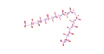

PEG-MGF, or Pegylated Mechano Growth Factor, is a variant of IGF-1. PEG-ylation modifies MGF by fusing polyethylene glycol (PEG) into it, potentially extending its half-life from a few minutes to days, allowing it to travel through the bloodstream for an extended period before breaking down and being excreted by the kidneys. Researchers suggest this process is essential for animal muscle regeneration after physical exertion or injury by promoting nitrogen retention and increasing protein synthesis.
Mechanism of Action
Research data from animal studies suggests that PEG-MGF supports the regeneration process of muscle, positively regulates protein synthesis, and activates satellite cells. In experiments where the PEG-MGF cDNA has been inserted into a plasmid vector and introduced into muscle cells, it appears to be a potent inducer of muscle hypertrophy. The addition of PEG-MGF to muscle myoblasts appeared to have increased proliferation and delayed differentiation, even in the presence of anti – IGF 1 receptor antibodies, potentially by activating fibrinolysis of matrix and metalloproteinase systems. Additionally, researchers propose that intense physical exertion may stimulate the release of growth hormone in the muscles to release MGF, especially as levels of this hormone decrease over time. Furthermore, a recent study of PEG-MGF suggests that the activity of protein kinase C is speculated to be necessary for the activation of this peptide (translocation to the nucleus) of factor 2 related to NFE 2 (Nrf2). This, in turn, is suggested to increase the expression of heme oxygenase 1, an event speculated to mediate neuroprotection of neurons from oxidative stress-induced apoptosis in the brain.
Research into PEG-MGF
In addition to its potential effects on muscle, PEG-MGF is speculated to have other impacts in the bodies of animal research models. Researchers suggest that PEG-MGF may enhance the proliferation and migration of bone marrow-derived mesenchymal stem cells, which are suggested to be a source of autologous stem cells for transplantation to the heart. There is speculative evidence of transient regulation of PEG b MGF expression in response to myocardial infarction associated with ischemia in the heart. Intracoronary exposure to this peptide is suggested to induce myocardial protection and improve hemodynamic function more than mature IGF – 1 after myocardial infarction in sheep. The speculated cellular protection conferred by PEG-MGF is suggested to be based on the inhibition of apoptosis in the border area of the infarct. PEG-MGF has been suggested to stimulate proangiogenic activities in vascular endothelial cells. Therefore, it may confer potentially beneficial action at the level of vascular regeneration and collateralization to restore blood flow to the heart after myocardial infarction. IGF – 1, considered to play an essential role in the interface between neurons in injured or damaged muscles, has shown potential effectiveness in slowing the progression of amyotrophic lateral sclerosis (ALS), a disease characterized by the loss of motor neurons and progressive muscle weakness. Exposure to PEG-MGF has been reported to produce improvements, with more motor neurons surviving in PEG-MGF-treated mice. MGF may be expressed in excess in regenerating regions after global cerebral ischemia. Its transcripts, thought to be expressed during brain development, are suggested to exhibit particular time distributions. Additionally, neonatal hypoxia and insults of hypoxic ischemia are suggested to lead to increased and prolonged expression of only the MGF isoform.
Disclaimer: The products mentioned are not intended for human or animal consumption. Research chemicals are intended solely for laboratory experimentation and/or in-vitro testing. Bodily introduction of any sort is strictly prohibited by law. All purchases are limited to licensed researchers and/or qualified professionals. All information shared in this article is for educational purposes only.