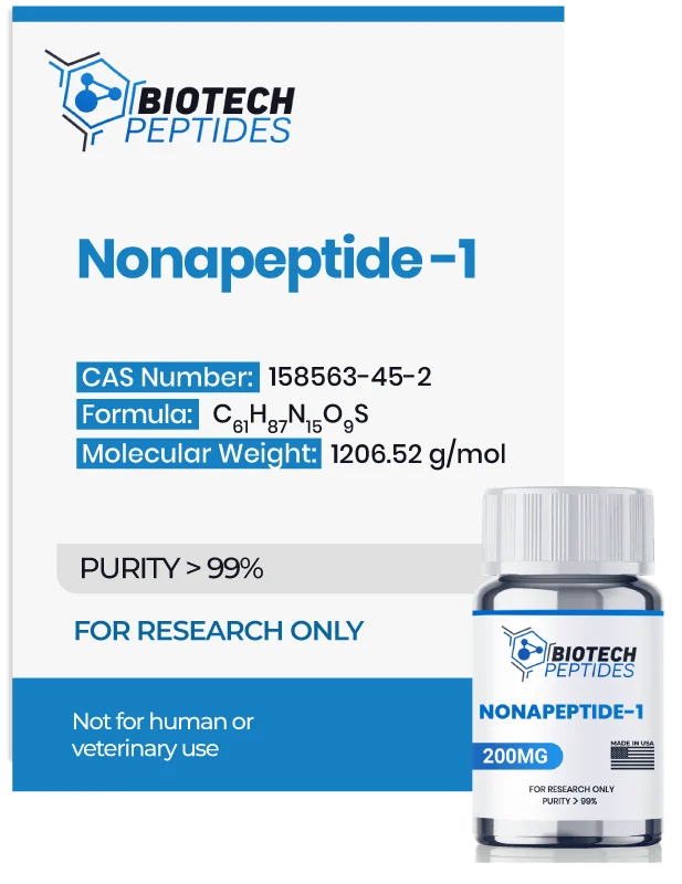
Nonapeptide-1 (200mg)
Original price was: $228.00.$219.00Current price is: $219.00.
Nonapeptide-1 peptides are Synthesized and Lyophilized in the USA.
Discount per Quantity
| Quantity | 5 - 9 | 10 + |
|---|---|---|
| Discount | 5% | 10% |
| Price | $208.05 | $197.10 |
FREE - USPS priority shipping
Nonapeptide-1 Peptide
Nonapeptide-1, also termed Melanostatine-5, is a peptide that may prevent the activation of pigment-producing cells in the skin called melanocytes. It appears to be one of the most potent inhibitors of a receptor found in these pigment-producing cells, called the melanocortin-1 receptor (MC1R). Researchers have explored a peptide library containing 31,360 structurally different compounds, and it appears that the most potent inhibitor identified was Met-Pro-D-Phe-Arg-D-Trp-Phe-Lys-Pro-Val-NH₂, aka Nonapeptide-1.[1] Structural analysis has revealed that specific amino acids, such as D-Trp at position 5 and Phe at position 6, are considered crucial for their antagonistic potential. The presence of D-Phe at position 3 further underscores such potential. Research on animal models suggests that Nonapeptide-1 may inhibit the synthesis of melanin, bringing it to the forefront of research on conditions impacting skin pigmentation, such as melasma. Studies in animal models posit that Nonapeptide-1 may reduce the synthesis of melanin and potentially decrease pigmentation on a considerable scale.[2]
Specifications
Sequence: Met-Pro-D-Phe-Arg-D-Trp-Phe-Lys-Pro-Val
Molecular Formula: C61H87N15O9S
Molecular Weight: 1206.52g/mol
Synonyms: Melanostatine, Oxytocin Intermediate-nine peptide, Melanostatine-5
Nonapeptide-1 Research
Nonapeptide-1 and General Мechanisms
Emerging research suggests that Nonapeptide-1 might decrease melanin production through multiple pathways. One potential mechanism may involve its interaction with the melanocortin 1 receptor (MC1R) found in melanocytes—the skin cells responsible for melanin synthesis. By binding to MC1R, Nonapeptide-1 may inhibit alpha-melanocyte-stimulating hormone (α-MSH) from activating this receptor. Normally, α-MSH, which appears to be a neuropeptide produced by the pituitary gland and skin cells, is posited to bind to MC1R and trigger a signaling cascade that activates an enzyme called tyrosinase, leading to increased melanin production. This process may increase skin pigmentation and may offer additional protection against ultraviolet (UV) radiation in normal settings. Thus, by blocking the activation of MC1R by α-MSH, Nonapeptide-1 could disrupt the signaling pathway that typically results in elevated tyrosinase activity and melanin synthesis in experimental models of dysregulated melanin synthesis.
In vitro studies using HaCaT cells (a keratinocyte cell line) and epidermal melanocytes (HEM) exposed to UV radiation indicate that Nonapeptide-1 may also reduce the expression levels of MC1R itself. This downregulation might further diminish the responsiveness of melanocytes to α-MSH. Furthermore, Nonapeptide-1 appears to significantly lower the expression of key proteins involved in melanin production, including tyrosinase-related proteins 1 and 2 (TRP1 and TRP2) and microphthalmia-associated transcription factor (MITF). Consequently, cells exposed to Nonapeptide-1 might exhibit a reduced capacity to increase melanin synthesis, even when exposed to UVA radiation.
Nonapeptide-1 and Skin Cell Pigmentation
Recent studies have explored nonapeptide-1 as a possible research compound, specifically in studies examining skin lightening in experimental models. Such studies have hypothesized that the compound might reduce skin pigmentation by approximately 33%, with the potential for ongoing improvement over time.[4] The main investigation into this peptide's potential was a prospective, double-blind, parallel-group, randomized controlled pilot study lasting eight months and divided into three phases.[5] Researchers observed what seemed to be improvements in the severity scores of melasma models— experimental models of a condition characterized by dark, discolored patches on the skin—and in the mean melanin index, which quantifies skin pigmentation levels. They noted that "the melasma area and severity index score showed a consistent reduction in the case group, whereas it increased in the control group from baseline," suggesting a positive potential of Nonapeptide-1 in managing experimental models of melasma.
Nonapeptide-1 and Tumor Cells
Research indicates that melanocortin 1 receptors (MC1R) may play a role in the growth and survival of specific types of tumor cells in experimental conditions, more specifically melanoma cells.[6] Mutations in the MC1R gene might be linked to variations in the risk of the occurrence of melanoma cells. In one study, scientists inhibited MC1R using natural inhibitors similar to Nonapeptide-1, which appeared to reduce melanin synthesis and decrease morphological heterogeneity (variations in cell shape and size) in murine B16-F10 melanoma cells. This inhibition seemed to result in slower tumor cell growth. The findings suggest that MC1R could be significant in regulating melanoma growth and morphology and that persistent inhibition of these receptors might significantly slow down the proliferation rate of tumor cells expressing them. It is important to note, however, that Nonapeptide-1 has yet to be specifically studied for its potential on melanoma tumor cells. Further research would be necessary to determine any impact Nonapeptide-1 might have in this context.
Nonapeptide-1 and Neurotransmitter Interactions
Scientific research posits that Nonapeptide-1 may function in the central nervous system at the dopaminergic and opioid receptors.[7] Specifically, melanocortin 1 receptors (MC1Rs) may be present in the periaqueductal gray (PAG) matter, a region in the midbrain considered to play a crucial role in pain perception and modulation, a process known as nociception.[8] In one notable study, researchers have compared genetically modified murine models to overproduce an endogenous antagonist of MC1R with unaltered control murine models.[9] They evaluated the mice's responses to various stimuli, both painful and non-painful, and examined their reactions to induced inflammatory pain and neuropathic pain. Additionally, the scientists assessed the mice's aversion to capsaicin—the active component in chili peppers known to activate the TRPV1 receptor, which responds to harmful heat sensations—using a behavioral test called the paired preference paradigm. This test measures preference or aversion by offering the animal models two choices and observing their selection behavior. The researchers observed that murine models overexpressing the MC1R antagonist appeared to exhibit a reduced response to inflammatory pain and a slower development of hypersensitivity and allodynia. These murine models also seemed to show less avoidance of moderate concentrations of capsaicin. Although similar research has not been conducted specifically with Nonapeptide-1 acting as an antagonist of MC1R, Nonapeptide-1 might produce comparable actions due to potential similarities in how it might interact with these receptors.
Disclaimer: The products mentioned are not intended for human or animal consumption. Research chemicals are intended solely for laboratory experimentation and/or in-vitro testing. Bodily introduction of any sort is strictly prohibited by law. All purchases are limited to licensed researchers and/or qualified professionals. All information shared in this article is for educational purposes only.
References
- Jayawickreme, C. K., Quillan, J. M., Graminski, G. F., & Lerner, M. R. (1994). Discovery and structure-function analysis of alpha-melanocyte-stimulating hormone antagonists. Journal of Biological Chemistry, 269(47), 29846-29854.
- Boo YC. Up- or Downregulation of Melanin Synthesis Using Amino Acids, Peptides, and Their Analogs. Biomedicines. 2020 Sep 1;8(9):322. doi: 10.3390/biomedicines8090322. PMID: 32882959; PMCID: PMC7555855.
- Chen J, Li H, Liang B, Zhu H. Effects of tea polyphenols on UVA-induced melanogenesis via inhibition of α-MSH-MC1R signalling pathway. Postepy Dermatol Alergol. 2022 Apr;39(2):327-335. doi: 10.5114/ada.2022.115890. Epub 2022 May 9. PMID: 35645678; PMCID: PMC9131962.
- Mohammed, Y. H., Moghimi, H. R., Yousef, S. A., Chandrasekaran, N. C., Bibi, C. R., Sukumar, S. C., Grice, J. E., Sakran, W., & Roberts, M. S. (2017). Efficacy, Safety and Targets in Topical and Transdermal Active and Excipient Delivery. Percutaneous Penetration Enhancers Drug Penetration Into/Through the Skin: Methodology and General Considerations, 369–391. https://doi.org/10.1007/978-3-662-53270-6_23
- Chatterjee, M., Neema, S., & Rajput, G. R. (2021). A randomized controlled pilot study of a proprietary combination versus sunscreen in melasma maintenance. Indian journal of dermatology, venereology and leprology, 88(1), 51–58. https://doi.org/10.25259/IJDVL_976_18
- Kansal, R. G., McCravy, M. S., Basham, J. H., Earl, J. A., McMurray, S. L., Starner, C. J., Whitt, M. A., & Albritton, L. M. (2016). Inhibition of melanocortin 1 receptor slows melanoma growth, reduces tumor heterogeneity and increases survival. Oncotarget, 7(18), 26331–26345. https://doi.org/10.18632/oncotarget.8372
- Puciłowski O, Płaźnik A, Kostowski W. Melanostatyna (MIF-1): działania ośrodkowe i próby kliniczne [Melanostatin (MIF-1): central action and clinical use]. Pol Tyg Lek. 1983 Jun 13;38(24):739-41. Polish. PMID: 6139794.
- Xia, Y., Wikberg, J. E., & Chhajlani, V. (1995). Expression of melanocortin 1 receptor in periaqueductal gray matter. Neuroreport, 6(16), 2193-2196.
- Delaney, A., Keighren, M., Fleetwood-Walker, S. M., & Jackson, I. J. (2010). Involvement of the melanocortin-1 receptor in acute pain and pain of inflammatory but not neuropathic origin. PloS one, 5(9), e12498. https://doi.org/10.1371/journal.pone.0012498


