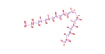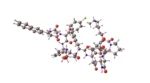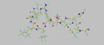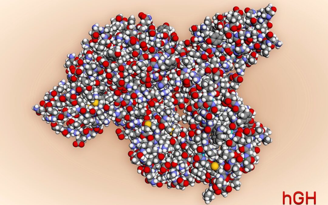
Frag 176-191 and Growth Hormone Interaction
Research
1. Frag 176-191 and Blood Sugar Levels:
Research suggests that Frag 176-191 is a potentially potent synthetic equivalent of the growth hormone and may act to reduce blood sugar levels. This potential is secondary to the increase in plasma insulin levels. Research is ongoing to determine if Frag 176-191 may be employed effectively in research studies using research models of prediabetes and type II diabetes.
2. Fragment 176-191 and Cartilage:
Though interest in Frag 176-191 is primarily researched for its potential its lipolytic functions, research is ongoing to determine its other possible impacts. In 2015, research from Korea suggested that Frag 176-191 increases the effects of hyaluronic acid exposure that may promote cartilage regeneration. Studies in rabbits suggested that Frag 176-191 increased cartilage growth laboratory standards, and its combination with hyaluronic acid may produce a more potent effect. The overall result suggests that exposure to Frag 176-191 alone and alongside hyaluronic acid might reduce the disabilities following osteoarthritis.
3. Frag 176-191 and Weight:
Fragment 176-191 is commonly referred to as Lipolytic Fragment because of its potential lipolytic potential. This function may possibly be mediated by increased beta-3 adrenergic receptor production (𝛃3-AR or ADRB3). The agonist action of ADRB3 is to accelerate fat burning in fatty tissues and thermogenesis in skeletal muscles. Mouse models genetically induced to not produce ADRB3 appear not to respond to the lipolytic potential of hGH or Frag 176-191. Research suggests that Frag 176-191 action may be accompanied by energy expenditure and weight reduction, amounting to about 50% of reduced weight gain in animal models of obesity over exposure for three weeks. Notably, the weight reduction effect was reported only in obese mice. In lean mice, they appeared to maintain their standard body weight even on exposure to Fragment 176-191. These results suggest that specific lipolysis pathways may be responsible for overriding the effect of ADRB3.
Disclaimer: The products mentioned are not intended for human or animal consumption. Research chemicals are intended solely for laboratory experimentation and/or in-vitro testing. Bodily introduction of any sort is strictly prohibited by law. All purchases are limited to licensed researchers and/or qualified professionals. All information shared in this article is for educational purposes only.