Thymosin Alpha 1: General Research and Scientific Observations

Thymosin Alpha 1: General Research and Scientific Observations

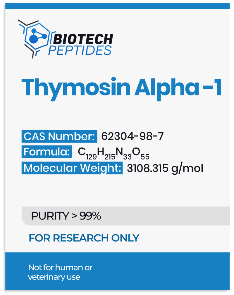
Thymosin Alpha 1 (Tα1) is a naturally occurring peptide initially isolated from thymus. It comprises 28 amino acids manifesting varied biological potential. These are considered to be highly diverse, including immune response augmentation, strengthening vaccine efficacy, and overcoming chemotherapy-induced and other toxicities. Besides this, the peptide may be relevant in research related to cancers like renal, hepatocellular, and non-small cell lung cancer. Scientific data has examined Ta1 in infectious and non-infectious ailments like psoriatic arthritis, cell aging, immunodeficiency (HIV), among others.

Regarding the isolation, purification, and preparation of Thymosin Alpha 1, it was initially isolated from the calf thymus in 1977. However, a solid-phase synthesis technique in which molecules are covalently bound to a solid material is predominantly used for its commercial production. With the arrival of molecular expression methods, Ta1 is also being produced through prokaryotic and eukaryotic expression methodologies.


Thymosin Alpha 1 Overview

The synthetic analog of Thymosin Alpha 1, namely Thymalfasin, is primarily studied in research models of hepatitis B and C infection. It has the following amino acid sequence:

Ac–S–D–A–A–V–Asp–T–S–S–E–I–T–T–K–D–L–K–E–K–K–E–V–V–E–E–A–E–N–OH.

As described above and shown in the sequence, the peptide is N-terminally acetylated, has a net positive charge, and lacks stable conformation.

Several research institutions and teams have aimed to explore the breadth of the peptide’s potential. However, the mechanisms by which this peptide exerts its effects are an active area of investigation. Scientists believe that Thymosin Alpha 1’s potential in overcoming infections, cancer, immunodeficiency, and cell aging, may be through pleiotropic action. A recent study suggested that Thymosin Alpha 1 interactions with an oligosaccharide binding protein, Galectin -1 (Gal-1), are necessary to activate its potential [1].

The immunostimulatory effects of Thymosin Alpha-1 peptide are supposed to be mediated through Toll-like receptors (TLRs). It has been observed that Thymosin Alpha 1, through its binding with TLR3/4/9, appears to stimulate interferon regulatory factor (IRF3) and NF-kB signaling pathways. The ultimate effect is reflected in the immune-enhancing potential for both innate and adaptive immune system machinery. The immunomodulation against viral infections also appears to be mediated through activating T cells, B cells, macrophages, and Natural Killer cells [2].

The molecular mechanisms underlying the potential of Ta1 in controlling cellular proliferation through inducing apoptosis of cancerous cells are also being evaluated. Scientific studies suggest that the hypothetical anti-cancer action of Thymosin Alpha 1 may be mediated through the PI3K/Akt/mTOR signaling pathway. Furthermore, PTEN appears to mediate apoptosis through Thymosin Alpha 1 [3]. The specificity of this is yet to be deciphered through further studies.

It is clear from the above-described studies that researchers general assert that Thymosin Alpha 1 appears to exert action through differential molecular and cellular mechanisms that should be investigated in further detail.


Thymosin Alpha 1 Peptide Research

Scientific studies have provided ample data to support its hypothetical consequence within the context of various ailments.

The primary objectives of several ongoing research studies are to evaluate this peptide’s potential individually or in combination with other compounds. Mainly, its action as a sole agent in the research of immune reconstitution disorders, hepatocellular carcinoma, osteoporotic pain, rheumatic heart disease, sepsis, and viral infections, is an active area of scientific investigation. The peptide, in combination with other compounds, is being evaluated for advanced refractory solid tumors, esophageal cancer, rectal carcinoma, malignant melanoma, and non-small cell lung cancer (NSCLC).

Besides prospective well-designed studies, several retrospective evaluations hypothesize the relevance of this peptide in various avenues of scientific research. Particularly in sepsis, organ dysfunction is a major focus. A study compared the benefits of Thymosin Alpha 1 with a placebo. The outcome revealed that Ta1 reduces organ damage among research models of sepsis [4].

Another study evaluated the potential of Thymosin Alpha 1 on long-term survival in margin-free (R0)-resected stage IA–IIIA with non-small cell lung cancer (NSCLC) models. Findings suggested that Thymosin Alpha 1 exposure appeared to have improved disease-free survival after R0 resection. The period of the study spanned 24 months [5].

The United States Patent Application Publication US 2010/0004174 A1 published in the year 2010 claimed that Thymosin Alpha 1 may exhibit potential benefits in cases of multiple sclerosis, inflammatory bowel disease, Crohn’s disease/ulcerative colitis.


Conclusion

Thymosin Alpha 1 is an endogenous peptide, whose synthetic analog, Thymalfasin, is widely researched as a prospective agent in various ailments. Most prominent theorized actions of Thymosin Alpha 1 are speculated to encompass tumor growth and cancer suppression, boosting immune cell functions, mitigating bacterial, viral, and fungal infections, acting as an anti-inflammatory agent, and augmenting vaccine activity. The most plausible explanation for its diverse potential is that Thymosin Alpha 1 may mediate homeostasis within different organs and systems. However, diverse mechanisms operate to control different diseases. Though a broader profile, this peptide molecular mechanism still needs to be elucidated for each proposed indication.


Disclaimer: The products mentioned are not intended for human or animal consumption. Research chemicals are intended solely for laboratory experimentation and/or in-vitro testing.  Bodily introduction of any sort is strictly prohibited by law.  All purchases are limited to licensed researchers and/or qualified professionals. All information shared in this article is for educational purposes only.


References

  1. Matteucci C, Nepravishta R, Argaw-Denboba A, Mandaliti W, Giovinazzo A, Petrone V, Balestrieri E, Sinibaldi-Vallebona P, Pica F, Paci M, Garaci E. Thymosin alpha1 interacts with Galectin-1 modulating the beta-galactosides affinity and inducing alteration in the biological activity. Int Immunopharmacol. May 2023;118:110113. doi:10.1016/j.intimp.2023.110113. PMID: 37028279. https://pubmed.ncbi.nlm.nih.gov/37028279/
  2. Tao N, Xu X, Ying Y, Hu S, Sun Q, Lv G, Gao J. Thymosin alpha1 and Its Role in Viral Infectious Diseases: The Mechanism and Clinical Application. Molecules. Apr 17 2023;28(8)doi:10.3390/molecules28083539. PMID: 37110771; PMCID: PMC10144173. https://pubmed.ncbi.nlm.nih.gov/37110771/
  3. Guo Y, Chang H, Li J, Xu XY, Shen L, Yu ZB, Liu WC. Thymosin alpha 1 suppresses proliferation and induces apoptosis in breast cancer cells through PTEN-mediated inhibition of PI3K/Akt/mTOR signaling pathway. Apoptosis. Aug 2015;20(8):1109-21. doi:10.1007/s10495-015-1138-9. PMID: 26002438. https://pubmed.ncbi.nlm.nih.gov/26002438/
  4. Cao M, Wang G, Xie J. Immune dysregulation in sepsis: experiences, lessons and perspectives. Cell Death Discov. Dec 19 2023;9(1):465. doi:10.1038/s41420-023-01766-7. PMID: 38114466; PMCID: PMC10730904. https://pubmed.ncbi.nlm.nih.gov/38114466/
  5. Guo CL, Mei JD, Jia YL, Gan FY, Tang YD, Liu CW, Zeng Z, Yang ZY, Deng SY, Sun X, Liu LX. Impact of thymosin alpha1 as an immunomodulatory therapy on long-term survival of non-small cell lung cancer patients after R0 resection: a propensity score-matched analysis. Chin Med J (Engl). Nov 3 2021;134(22):2700-2709. doi:10.1097/CM9.0000000000001819. PMID: 34732663; PMCID: PMC8631386. https://www.ncbi.nlm.nih.gov/pmc/articles/PMC8631386/
Tesamorelin & Modified GRF 1-29, & Ipamorelin Peptide Blend – Potential Synergism

Tesamorelin & Modified GRF 1-29, & Ipamorelin Peptide Blend – Potential Synergism

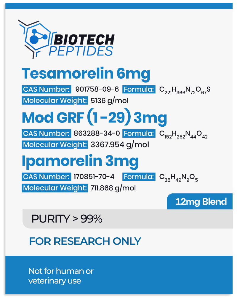
Tesamorelin, Modified GRF 1-29, and Ipamorelin represent a cohort of synthetic peptides with disparate mechanisms, converging on the probable stimulation of growth hormone (GH) synthesis and secretion.

Tesamorelin, functioning as a synthetic analogue of growth hormone-releasing hormone (GHRH), appears to exhibit a distinctive potential to elicit the production and release of endogenous growth hormone (GH). Empirical investigations indicate a discernible selectivity in activating GH-releasing hormone receptors within the pituitary gland. This selectivity augments its efficacy in manifesting the purported physiological effects.[1]

In the case of Modified GRF 1-29, this synthetic peptide manifests an extended half-life, likely attributable to its resistance to enzymatic degradation. It has been hypothesized to operate as a robust stimulator of GH secretion, showcasing considerable potential in fostering protein synthesis, facilitating muscle cell growth, and enhancing metabolic processes.[2]

Finally, Ipamorelin, a synthetic pentapeptide, has been suggested by researchers to operate as a selective agonist for the ghrelin receptor, thereby instigating the release of GH. Its noteworthy potential lies in a high specificity and minimal impact on other hormonal systems, positioning it as a valuable research instrument for precision-targeted GH stimulation.[3]  This specific attribute renders Ipamorelin an invaluable tool for scientific inquiries directed at understanding and manipulating GH dynamics.


Tesamorelin, Modified GRF and Ipamorelin Blend and Pituitary Gland

The synergy of Tesamorelin, Mod GRF 1-29, and Ipamorelin has been suggested through their substantive interactions with the pituitary gland. These interactions are characterized by precise receptor binding, leading to the modulation of growth hormone (GH) release.

Tesamorelin appears to exhibit an affinity for binding predominantly to growth hormone-releasing hormone (GHRH) receptors located on the surface of somatotrophs in the anterior pituitary gland. Upon this binding, Tesamorelin is speculated to initiate the activation of adenylate cyclase, thereby initiating the production of cyclic adenosine monophosphate (cAMP). This series of molecular events appears to carry the potential to induce the release of growth hormone (GH).[4] Meanwhile, Modified GRF, apparently engaging with GHRH receptors, emerges as a potential GH secretagogue. Its interaction with GHRH receptors is said to enhance GH release, concomitantly promoting protein synthesis, facilitating muscle cell growth, and amplifying metabolic processes—a phenomenon supported by speculative scientific research.

The structural configuration of the Modified GRF peptide, marked by four amino acid substitutions, appears to enhance its GH-related activity and fortifies its resistance against proteolytic enzymes. These structural modifications are purported to facilitate the covalent binding of “at least 90% of the peptide” to blood albumin, with trace amounts potentially binding to fibrinogen and immunoglobulin G (IgG).

Researchers report “No other chemical species have been found bound to DAC-GRF after [introduction]. This binding extends the half-life of the active pharmacophore, resulting in a markedly prolonged duration of action in several animal species.”[5]

In contrast, Ipamorelin appears to act as a selective agonist for the ghrelin receptor, which is also expressed on somatotrophs within the pituitary gland. Its binding to ghrelin receptors is posited to induce GH release with high specificity and minimal impact on other hormonal systems. Research findings underscore that Ipamorelin exposure may lead to heightened GH secretion without markedly affecting cortisol, prolactin, or insulin levels, thereby offering a prospect of more targeted and controlled GH stimulation.


Tesamorelin, Mod GRF 1-29 and Ipamorelin Blend and Gastrointestinal Tract

Within the trio of growth hormone-releasing peptides, Tesamorelin has been suggested by researchers to display an apparent capacity to augment gastric emptying and enhance gastrointestinal motility in animal models. Meanwhile, Modified GRF, while exhibiting a minimal impact, appears to contribute to the improvement of gut barrier function and the reduction of intestinal inflammation in animal research models of colitis. Ipamorelin is posited to predominantly interact with the ghrelin receptor within the gastrointestinal (GI) tract.

Following binding to the ghrelin receptors, Ipamorelin may evoke diverse responses, including the promotion of gut motility and the improvement of intestinal absorption. Additionally, Ipamorelin exhibits promise in mitigating inflammation and fostering tissue repair across various models of GI injury. Researchers assert that Ipamorelin may “increase total body fat percentages,” designating the peptide as a “potent and selective stimulator of GH that [may] significantly influence the GI system, body composition, and adiposity.”[7]


Tesamorelin, Modified GRF and Ipamorelin Blend and Cardiovascular System

Myocardial Infarction (MI) is recognized for inducing scarring in cardiac tissues, potentially compromising heart function, including parameters like ejection fraction.

Animal model studies suggest that growth hormone may expedite the repair of heart tissues post-MI, leading to a reduction in infarct size, enhancement of cardiac ejection fraction, and an overall improvement in cardiac function.[8]

Researchers observe that, “Through activation of GHS-R1a, secretagogues produced a positive inotropic effect on ischemic cardiomyocytes and protected them from I/R injury, likely by safeguarding or restoring p-PLB (and hence SR Ca2+ content) to facilitate the maintenance or recovery of normal cardiac contractility.”

Research underscores that Tesamorelin, beyond its potential in reducing lipodystrophy, may also contribute to notable reductions in triglyceride levels, total cholesterol levels, and non-HDL-C levels, especially among HIV-positive models.[9]


Synergistic Potential of Tesamorelin, Mod GRF 1-29 and Ipamorelin Blend

The amalgamation of these peptides is underpinned by the rationale rooted in their distinct mechanisms of action and the potential for synergistically enhanced growth hormone (GH) secretion.

An experimental study elucidated a synergistic augmentation in GH secretion when Tesamorelin and Modified GRF were combined, surpassing the effects observed with either peptide introduced individually. This synergistic hypothesis is attributed to the complementary mechanisms of action and receptor interactions inherent to these peptides.[6]

Notably, in research models of HIV-associated lipodystrophy, a combination of Tesamorelin and Ipamorelin exhibited more pronounced reductions in visceral adipose tissue compared to Tesamorelin in isolation. However, researchers caution that this effect is transient and reversible upon cessation of exposure.

Scientific research has documented diverse potential actions associated with this peptide blend, encompassing accelerated fat loss, improved sleep quality, mood enhancement, increased energy levels, and potential sustained long-term growth hormone (GH) production, particularly purported with the combined exposure of Ipamorelin and Modified GRF. The protracted elevation of GH production post-cycle may contribute to the enduring maintenance of these benefits over an extended period.

Tesamorelin assumes a pivotal role in enhancing the overall physiological framework of the body. Among the trio of growth hormone-releasing peptides, Tesamorelin is esteemed for its versatility. The concurrent introduction of these three peptides appears to yield a synergistic impact on GH secretion and insulin-like growth factor 1 (IGF-1) levels. This potential synergy may lead to heightened GH secretion, augmented muscle cell growth, improved bone density, cognitive enhancement, cellular repair and regeneration, fat loss, and other multifaceted physiological impacts.


Disclaimer: The products mentioned are not intended for human or animal consumption. Research chemicals are intended solely for laboratory experimentation and/or in-vitro testing.  Bodily introduction of any sort is strictly prohibited by law.  All purchases are limited to licensed researchers and/or qualified professionals. All information shared in this article is for educational purposes only.


References

  1. National Center for Biotechnology Information (2023). PubChem Compound Summary for CID 9831659, Ipamorelin. https://pubchem.ncbi.nlm.nih.gov/compound/Ipamorelin.
  2. National Center for Biotechnology Information (2023). PubChem Compound Summary for CID 56841945. https://pubchem.ncbi.nlm.nih.gov/compound/56841945.
  3. National Center for Biotechnology Information (2023). PubChem Compound Summary for CID 16137828, Tesamorelin. https://pubchem.ncbi.nlm.nih.gov/compound/Tesamorelin.
  4. Clinical and Research Information on Drug-Induced Liver Injury [Internet]. Bethesda (MD): National Institute of Diabetes and Digestive and Kidney Diseases; 2012-. Tesamorelin. [Updated 2018 Oct 20]. https://www.ncbi.nlm.nih.gov/books/NBK548730/
  5. Sam L. Teichman and others, Prolonged Stimulation of Growth Hormone (GH) and Insulin-Like Growth Factor I Secretion by CJC-1295, a Long-Acting Analog of GH-Releasing Hormone, in Healthy Adults, The Journal of Clinical Endocrinology & Metabolism, Volume 91, Issue 3, 1 March 2006, Pages 799–805, https://doi.org/10.1210/jc.2005-1536 ; https://academic.oup.com/jcem/article/91/3/799/2843281
  6. Bedimo R. Growth hormone and tesamorelin in the management of HIV-associated lipodystrophy. HIV AIDS (Auckl). 2011;3:69-79. doi: 10.2147/HIV.S14561. Epub 2011 Jul 10. PMID: 22096409; PMCID: PMC3218714. https://www.ncbi.nlm.nih.gov/pmc/articles/PMC3218714/
  7. Sinha DK, Balasubramanian A, Tatem AJ, Rivera-Mirabal J, Yu J, Kovac J, Pastuszak AW, Lipshultz LI. Beyond the androgen receptor: the role of growth hormone secretagogues in the modern management of body composition in hypogonadal males. Transl Androl Urol. 2020 Mar;9(Suppl 2):S149-S159. doi: 10.21037/tau.2019.11.30. PMID: 32257855; PMCID: PMC7108996 https://www.ncbi.nlm.nih.gov/pmc/articles/PMC7108996/
  8. Ma Y, Zhang L, Edwards JN, Launikonis BS, Chen C. Growth hormone secretagogues protect mouse cardiomyocytes from in vitro ischemia/reperfusion injury through regulation of intracellular calcium. PLoS One. 2012;7(4):e35265. https://journals.plos.org/plosone/article?id=10.1371/journal.pone.0035265 Epub 2012 Apr 6. PMID: 22493744; PMCID: PMC3320867.
  9. Stanley TL, Falutz J, Marsolais C, Morin J, Soulban G, Mamputu JC, Assaad H, Turner R, Grinspoon SK. Reduction in visceral adiposity is associated with an improved metabolic profile in HIV-infected patients receiving tesamorelin. Clin Infect Dis. 2012 Jun;54(11):1642-51. doi: 10.1093/cid/cis251. Epub 2012 Apr 10. PMID: 22495074; PMCID: PMC3348954. https://pubmed.ncbi.nlm.nih.gov/22495074/
Semaglutide Peptide: Research in Diabetes and Glycemic Control

Semaglutide Peptide: Research in Diabetes and Glycemic Control

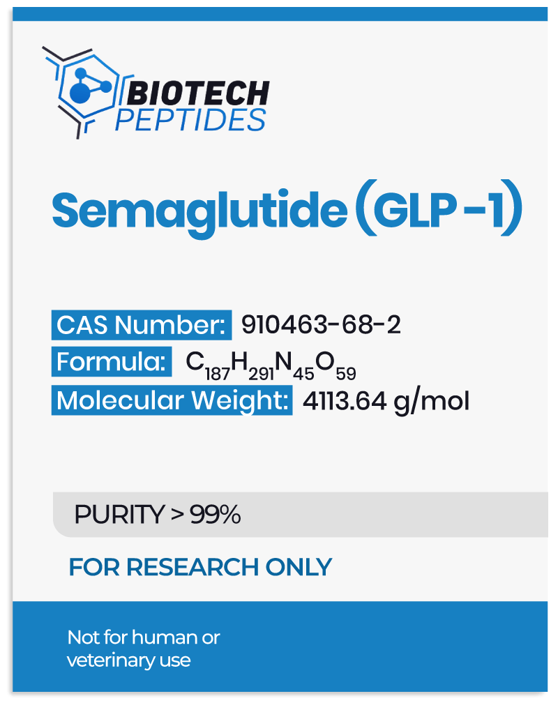
Semaglutide peptide is a novel glucagon-like peptide-1 (GLP-1) receptor agonist. It has emerged as an innovative compound within the realm of type 2 diabetes mellitus and obesity research. This article provides an overview of Semaglutide, its proposed mechanism of action, essential functions, historical development, and research data. Semaglutide peptide is a synthetic analog of glucagon-like peptide-1 (GLP-1). It is developed and refined through structural modifications (for enhanced stability and pharmacokinetic potential) to exert potential activity through the activation of the GLP-1 receptor.[1]

Semaglutide peptide is a long-acting GLP-1 receptor agonist that shares a structural similarity with native GLP-1. Researchers have suggested that the compound exhibits an improved resistance to degradation by dipeptidyl peptidase-4 (DPP-4). This modification is assumed to prolong its half-life.[2] Semaglutide  is presumed to activate the GLP-1 receptor on pancreatic beta cells by binding itself to it. This may prompt the insulin secretion in a glucose-dependent manner. Simultaneously, it appears to suppress glucagon secretion. Research suggests that this slows down gastric emptying, modulates appetite regulation centers, and contributes to glycemic control, potentially helping weight management.


Development of Semaglutide Peptide

The Semaglutide peptide precursor appears to play a vital role in the biosynthesis and maturation of the active peptide. The precursor is a pro-peptide derived from the glucagon-like peptide-1 (GLP-1) analog. It appears to undergo intricate processing within pancreatic cells, influencing the production and secretion of the bioactive Semaglutide molecule. The precursor peptide of Semaglutide is initially translated from the corresponding gene transcript. The post-translational modifications, including cleavage by prohormone convertases such as PC1/3, occur within pancreatic alpha cells. They are considered to facilitate the generation of the mature peptide.[5] This multi-step process involves the removal of signal peptides and the formation of disulfide bonds critical for structural stability.

The mature Semaglutide and its precursor undergo intricate intracellular trafficking mechanisms, after processing. They traverse the secretory pathway, involving the endoplasmic reticulum and Golgi apparatus. There, the proper folding, post-translational modifications, and packaging into secretory vesicles occur. The regulated secretion of the mature peptide involves complex cellular machinery to ensure appropriate release upon physiological stimuli. The Semaglutide precursor appears to serve as a template for the active peptide’s formation and may exert regulatory and modulatory influences within the secretory pathway. It may also participate in the quality control mechanisms. Therefore, researchers suggest, it may act to ensure correct folding and prevent premature degradation. Recent studies also suggest potential intracellular signaling roles of pro-peptides, influencing cellular processes beyond peptide maturation.[6]

The desire to harness the potential of glucagon-like peptide-1 (GLP-1) within the context of type 2 diabetes mellitus (T2DM) and obesity marks the beginning of Semaglutide research. The evolutionary journey of this peptide involves iterative modifications, all aimed at identifying and honing its physiological activity. The quest for GLP-1 analogs started with the recognition of native GLP-1’s potential impact on glucose regulation. However, the peptide’s reputed short half-life due to rapid degradation by dipeptidyl peptidase-4 (DPP-4) called for structural alterations to enhance stability while retaining its purported pharmacological activity. Semaglutide’s development involved meticulous structural modifications. These were mainly focused on alterations in amino acid sequence and attachment of fatty acid side chains. These adaptations aimed to ensure resistance to enzymatic degradation and extend its duration of action.[8]

Preclinical investigations involving cell culture models, animal studies, and pharmacokinetic assessments played a central role in refining Semaglutide’s properties. The findings of these studies first suggested the peptide’s action in improving glycemic control and inducing weight loss.


Research and Scientific Studies


Semaglutide Peptide and Diabetes

Numerous clinical trials have examined Semaglutide’s potential within type-2 diabetes mellitus research.[3] The study findings imply that Semaglutide peptide exhibits superior reductions in HbA1c levels compared to placebo and other antidiabetic agents. Moreover, its effectiveness may extend to obesity research, potentially leading to substantial weight loss in research models with and without diabetes.

The continued research of Semaglutide peptide centers on expanding the understanding of its mechanisms and exploring the potential research applications beyond type-2 diabetes mellitus and obesity. Additionally, researchers also focus on refining Semaglutide’s formulation for improved adherence and outcomes.


Semaglutide and Glycemic Control

Semaglutide peptide has primarily been researched for its potential to enhance glycemic control through glucose-dependent insulin secretion and suppression of glucagon release. Additionally, researchers have suggested its potential in improving insulin sensitivity in peripheral tissues, possibly contributing to better glucose utilization and mitigating insulin resistance.[15]


Semaglutide peptide and Weight, Satiety Control

Semaglutide peptide has been suggested to induce substantial weight loss in research models of type-2 diabetes mellitus, as well as models of obesity. It appears to delay gastric emptying, which may lead to prolonged satiety, and the modulation of appetite-regulating centers in the brain, which seem to collectively contribute to sustained weight reduction.

A study published in October 2022, examining the potential impact of Semaglutide peptide, included obese and overweight research models, with or without type-2 diabetes mellitus.[9] They were divided into two groups. One group received Semaglutide peptide once a week, while the other group received a placebo. The results of the study reported that 69% to 79% of non-diabetic research models in the Semaglutide group recorded an average weight loss of ≥10% by week 68, while 51% to 65% achieved a weight loss of ≥15%, compared to ≥5% – 13% in the placebo group. By week 104, the weight loss was 15.2% in the Semaglutide peptide group versus 2.6% in the placebo group. Obese and overweight models with type-2 diabetes mellitus recorded an average weight loss of 9.6% by week 68, versus 3.4% in the placebo group.

A double-blind, parallel-group study in 2021, suggested that Semaglutide peptide may have led to reduced hunger and prospective food consumption, while potentially also increasing fullness and satiety when compared with placebo.[14] A better control of eating and fewer food cravings were indicated with Semaglutide versus placebo. Body weight was reduced by 9.9% with Semaglutide and 0.4% with placebo.


Semaglutide peptide and Cardiovascular, Renal Function

Emerging research data suggests a potential for Semaglutide peptide to confer cardiovascular action. This may include reductions in cardiovascular events in high-risk cases. Moreover, ongoing research explores its potential influence on renal function. Some findings indicate potential renoprotective characteristics beyond the possible glycemic control.

A study from 2022 examining Semaglutide peptide on cardiometabolic risk factors in research models of obesity states that: “Semaglutide may improve cardiometabolic risk factors and reduce antihypertensive/lipid‐lowering [compounds] use versus placebo in [research subjects] with overweight/obesity without diabetes. These potential benefits were not maintained after … discontinuation”.[10]

Another study published in 2023 suggests that Semaglutide peptide may have improved urine albumin-to-creatinine ratio (UACR) in research models of obesity and type 2 diabetes, respectively.[11] At the same time, in control models, Semaglutide did not appear to affect eGFR decline.


Semaglutide Peptide and Neurology

Beyond its purported metabolic actions, Semaglutide peptide may exert some degree of neuroprotective potential. Researchers hint at its ability to traverse the blood-brain barrier, exerting neurotropic potential and displaying neuroprotective characteristics in models of neurodegenerative diseases.

A 2018 murine model suggests that Semaglutide peptide may potentially have an impact in Parkinson’s disease.[12] Both GLP-1 analogs, Semaglutide and Liraglutide, appeared to have improved 1-methyl-4-phenyl-1, 2, 3, 6-tetrahydropyridine (MPTP)-induced motor impairments in mice. In addition, both rescued the decrease of tyrosine hydroxylase (TH) levels, alleviated the inflammation response, reduced lipid peroxidation, inhibited the apoptosis pathway, and also increased autophagy-related protein expression, to protect dopaminergic neurons in the substantia nigra and striatum, as reported by the researchers.


Semaglutide and Inflammation, Metabolic Modulation

Semaglutide’s potential may extend to modulating inflammatory pathways, potentially exerting anti-inflammatory impact. It also appears to influence various metabolic pathways, impacting lipid metabolism, adipose tissue function, and hepatic steatosis. A 2022 study on mice suggests that the body weight, liver weight, blood glucose, TG, TCHO, LDL, and pro-inflammatory factors of animal subjects were significantly reduced after Semaglutide peptide exposure.[13] At the same time, Semaglutide appeared to have increased the SOD level. The exposure may have also significantly improved the pathological changes such as hepatocyte steatosis, balloon degeneration, and lymphoid foci by HE.

So far, the research results suggest that Semaglutide, a prominent GLP-1 receptor agonist, maybe a significant research innovation in the sphere of metabolic function studies. Its potentially robust activity in glycemic control, combined with possibly substantial weight reduction, may be a paradigm shift in type 2 diabetes mellitus and obesity research.


Disclaimer: The products mentioned are not intended for human or animal consumption. Research chemicals are intended solely for laboratory experimentation and/or in-vitro testing.  Bodily introduction of any sort is strictly prohibited by law.  All purchases are limited to licensed researchers and/or qualified professionals. All information shared in this article is for educational purposes only.


References:

  1. Andersen A, Knop FK, Vilsbøll T. A Pharmacological and Clinical Overview of Oral Semaglutide for the Treatment of Type 2 Diabetes. Drugs. 2021;81(9):1003-1030. doi: 10.1007/s40265-021-01499-w
  2. Mahapatra MK, Karuppasamy M, Sahoo BM. Semaglutide is a glucagon-like peptide-1 receptor agonist with cardiovascular benefits for the management of type 2 diabetes. Rev Endocr Metab Disord. 2022 Jun;23(3):521-539. doi: 10.1007/s11154-021-09699-1. Epub 2022 Jan 7. PMID: 34993760; PMCID: PMC8736331.
  3. Goldenberg RM, Steen O. Semaglutide: Review and Place in Therapy for Adults With Type 2 Diabetes. Can J Diabetes. 2019;43(2):136-145. doi: 10.1016/j.jcjd.2018.05.008
  4. Garvey WT, Batterham RL, Bhatta M, et al. Two-year effects of semaglutide in adults with overweight or obesity: the STEP 5 trial. Nat Med. 2022;28(10):2083-2091. doi: 10.1038/s41591-022-02026-4
  5. Lafferty RA, O’Harte FPM, Irwin N, Gault VA, Flatt PR. Proglucagon-Derived Peptides as Therapeutics. Front Endocrinol (Lausanne). 2021 May 18;12:689678. doi: 10.3389/fendo.2021.689678. PMID: 34093449; PMCID: PMC8171296.
  6. Cunha FM, Berti DA, Ferreira ZS, Klitzke CF, Markus RP, Ferro ES. Intracellular peptides as natural regulators of cell signaling. J Biol Chem. 2008 Sep 5;283(36):24448-59. doi: 10.1074/jbc.M801252200. Epub 2008 Jul 10. PMID: 18617518; PMCID: PMC3259820.
  7. Zhao X, Wang M, Wen Z, Lu Z, Cui L, Fu C, Xue H, Liu Y, Zhang Y. GLP-1 Receptor Agonists: Beyond Their Pancreatic Effects. Front Endocrinol (Lausanne). 2021 Aug 23;12:721135. doi: 10.3389/fendo.2021.721135. PMID: 34497589; PMCID: PMC8419463.
  8. Battle of GLP-1 delivery technologies – Scientific Figure on ResearchGate. Available from: https://www.researchgate.net/figure/A-Half-life-extension-mechanisms-of-acylated-GLP-1-analogs-B-Mean-concentration-time_fig2_326447155 [accessed 6 Dec 2023]
  9. Bergmann NC, Davies MJ, Lingvay I, Knop FK. Semaglutide for the treatment of overweight and obesity: A review. Diabetes Obes Metab. 2023;25(1):18-35. doi: 10.1111/dom.14863
  10. Kosiborod MN, Bhatta M, Davies M, Deanfield JE, Garvey WT, Khalid U, Kushner R, Rubino DM, Zeuthen N, Verma S. Semaglutide improves cardiometabolic risk factors in adults with overweight or obesity: STEP 1 and 4 exploratory analyses. Diabetes Obes Metab. 2023 Feb;25(2):468-478. doi: 10.1111/dom.14890. Epub 2022 Oct 28. PMID: 36200477; PMCID: PMC10092593.
  11. Heerspink HJL, Apperloo E, Davies M, Dicker D, Kandler K, Rosenstock J, Sørrig R, Lawson J, Zeuthen N, Cherney D. Effects of Semaglutide on Albuminuria and Kidney Function in People With Overweight or Obesity With or Without Type 2 Diabetes: Exploratory Analysis From the STEP 1, 2, and 3 Trials. Diabetes Care. 2023 Apr 1;46(4):801-810. doi: 10.2337/dc22-1889. PMID: 36801984; PMCID: PMC10090901.
  12. Zhang L, Zhang L, Li L, Hölscher C. Neuroprotective effects of the novel GLP-1 long-acting analog semaglutide in the MPTP Parkinson’s disease mouse model. Neuropeptides. 2018;71:70-80. doi: 10.1016/j.npep.2018.07.003
  13. Niu S, Chen S, Chen X, Ren Q, Yue L, Pan X, Zhao H, Li Z, Chen X. Semaglutide ameliorates metabolism and hepatic outcomes in an NAFLD mouse model. Front Endocrinol (Lausanne). 2022 Dec 9;13:1046130. doi: 10.3389/fendo.2022.1046130. PMID: 36568109; PMCID: PMC9780435.
  14. Friedrichsen M, Breitschaft A, Tadayon S, Wizert A, Skovgaard D. The effect of semaglutide 2.4 mg once weekly on energy intake, appetite, control of eating, and gastric emptying in adults with obesity. Diabetes Obes Metab. 2021;23(3):754-762. doi: 10.1111/dom.14280
  15. Andreadis P, Karagiannis T, Malandris K, et al. Semaglutide for type 2 diabetes mellitus: A systematic review and meta-analysis. Diabetes Obes Metab. 2018;20(9):2255-2263. doi: 10.1111/dom.13361
B7-33 Peptide: Research in Preeclampsia and Vasoprotection

B7-33 Peptide: Research in Preeclampsia and Vasoprotection

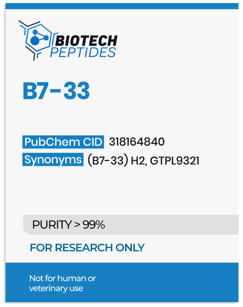
B7-33, a monomeric peptide, represents a synthetic derivation in soluble form from the endogenous protein H2-relaxin. This innovative peptide appears to retain the intrinsic anti-fibrotic attributes of relaxin, and researchers suggest it may strategically avoid the upregulation of cyclic adenosine monophosphate (cAMP) production. Beyond these foundational proposed characteristics, B7-33 appears to exhibit the unique potential to induce the phosphorylation of extracellular signal-regulated kinases 1 and 2 (ERK1/2) and enhance the expression of matrix metalloproteinase 2 (MMP2), possibly contributing to the degradation of extracellular collagen.[1]


Protein and Peptide Structural Elucidation

The H2-relaxin protein, a complex ensemble comprising relaxin, H3-relaxin, insulin-like peptide-3, and insulin-like peptide-5, is considered by scientists to govern diverse biological impacts, extending its influence to gene regulation and reproductive, musculoskeletal, and cardiovascular systems. This protein family appears to intricately interact with four distinct receptors – RXFP-1, RXFP-2, RXFP-3, and RXFP-4 – each wielding specific physiological actions.

RXFP-1, for instance, is considered to modulate sperm motility, joint function, and may play a role in pregnancy. RXFP-2 appears to influence testicular descent, RXFP-3 is implicated in sleep regulation, and RXFP-4 exhibits indications of involvement in hunger cycles. Given the expansive array of receptor interactions and the consequential biological impacts, intensive investigations have been undertaken, delving into the H2-relaxin protein and its derivatives, including the intriguing B7-33 peptide, to unveil their manifold research implications.


B7-33 Peptide Synthesis and Structural Modification

The conventional relaxin peptide encompasses four distinct components: a signal peptide, a B chain, a C chain, and a COOH terminal. Initial attempts to replicate this intricate structure resulted in high insolubility and inactivity in peptides. However, through meticulous structural modifications involving the production of a B chain and elongation of the COOH terminal, scientists achieved a groundbreaking milestone in 2016 – synthesizing the first-ever soluble analog, B7-33 peptide.[3]


B7-33 and Activation of pERK Pathway

Current research suggests that the B7-33 peptide may diverge from the canonical cAMP pathway typically associated with the anti-fibrotic properties of H2-relaxin. Activation of the cAMP pathway by H2-relaxin is considered to stimulate tumorigenesis, an adverse action of relaxin exposure. Intriguingly, the B7-33 peptide appears to display a notable affinity for RXFP-1 receptors. Binding the peptide to these receptors may induce pERK pathway activation, potentially resulting in heightened synthesis of matrix metalloproteinase 2 (MMP-2). The pivotal role of MMP-2 lies in inhibiting tissue scarring, thus providing a promising avenue for fibrosis research.[2]


Research and Scientific Studies


B7-33 Peptide and Preeclampsia

Preeclampsia is characterized by maternal hypertension and fetal growth restriction. A pivotal study delved into the impact of B7-33 peptide exposure on research models of preeclampsia using cytotrophoblasts. The results indicated an elevation in vascular endothelial growth factor (VEGF) levels, indicating the potential of B7-33 to counteract elevated glucose and marinobufagenin (MBG) levels, thereby attenuating the pathophysiology of preeclampsia.[4]

As per researchers Syeda H Afroze et al: ”Both B7-33 and its lipidated derivative mitigate the MBG- and hyperglycemia-induced dysfunction of CTBs by attenuating anti-angiogenic phenotype similar to that seen in preE. Moreover, the B7-33 and its lipidated derivative-induced effect on CTBs are attenuated by a relaxin antagonist.”[4]


B7-33 Peptide and Vasoprotection

H2-relaxin has earned its reputation as a vasoprotective compound among researchers, as a potential mitigator of cardiac failure and fibrosis. However, the cumbersome and expensive exogenous production of H2-relaxin prompted investigations into its analog, B7-33 peptide. In a comprehensive study involving male Wistar rats, vascular functions were meticulously assessed after the introduction of a placebo peptide, H2-relaxin, or B7-33 peptide. While impacts in the renal artery and abdominal aorta were negligible, both B7-33 and H2-relaxin appeared to have exhibited improvements in vasodilatory properties in the mesenteric artery. Another study in female mice hinted at potential effectiveness in preventing endothelial dysfunction induced by placental trophoblast conditioned media.[5]

Sarah A. Marshall et al. state, “In conclusion, … B7-33 replicated the acute beneficial vascular effects of serelaxin in rat mesenteric arteries and also prevented endothelial dysfunction induced by placental trophoblast conditioned media in mouse mesenteric arteries.”[5]


B7-33 Peptide and Anti-Fibrosis

Studies have posited that fully extended H2-relaxin may inadvertently stimulate carcinogenic cell proliferation via cAMP activation. In stark contrast, introduction of the B7-33 peptide to mice models of myocardial infarction indicated a substantial reduction in cardiac tissue fibrosis and improved heart function. This remarkable result was attributed to an increased concentration of matrix metalloproteinase protein, countering collagen-damaging cells and preventing fibrosis. Furthermore, in a prostate cancer study, both low and high concentrations of B7-33 appeared to have supported the prevention of fibrosis development and tumor spread. This finding suggested the exclusive activation of the pERK pathway, thereby possibly preventing cancer cell spread without activating cAMP. [6,7]


B7-33 Peptide as Coating Material

To surmount the immune response against foreign substances, a potential solution involves coating internal devices with the B7-33 peptide. A meticulous study in mice involved the implantation of a device coated with the peptide, which was released to counteract the fibrotic effects on the research models. The results indicated a significant reduction in device thickness induced by fibrosis over the 6-week duration of the study. While this marks only the initial step, these findings strongly suggest the potential of B7-33 peptide within the realm of coating material for devices and implants, though further research is ongoing.


In Summary

B7-33 peptide, a structural variant of H2-relaxin, emerges as a promising research compound with distinctive potential action. Its proposed action to activate the pERK pathway without inducing cAMP makes it a unique candidate in the realm of anti-fibrotic and vasoprotective research. The notable anti-fibrotic and vasoprotective potential of B7-33 signify its possible relevance in research related diverse cardiovascular and pulmonary disorders and preeclampsia.


Disclaimer: The products mentioned are not intended for human or animal consumption. Research chemicals are intended solely for laboratory experimentation and/or in-vitro testing.  Bodily introduction of any sort is strictly prohibited by law.  All purchases are limited to licensed researchers and/or qualified professionals. All information shared in this article is for educational purposes only.


References:

  1. Summers RJ. Recent progress in the understanding of relaxin family peptides and their receptors. Br J Pharmacol. 2017 May;174(10):915-920. doi: 10.1111/bph.13778. PMID: 28447360. https://www.ncbi.nlm.nih.gov/pmc/articles/PMC5406287/
  2. Mohammed Akhter Hossain et al., A single-chain derivative of the relaxin hormone is a functionally selective agonist of the G protein-coupled receptor, RXFP1, Drug Discovery Biology Pharmacology Monash Biomedicine Discovery Institute, Vol 7, 2016.
  3. Nitin A Patil et al, Relaxin family peptides: structure–activity relationship studies, British Pharmacological Society, vol 174 issue 10, published 06 December 2016. https://doi.org/10.1111/bph.13684
  4. H Afroze et al., Abstract P3042: Novel Peptide B7-33 and Its Lipidated Derivative Protect Cytotrophoblasts From Preeclampsia Phenotype in a Cellular Model of the Syndrome, 4 Sep 2019. https://doi.org/10.1161/hyp.74.suppl_1.P3042
  5. Marshall SA, O’Sullivan K, Ng HH, Bathgate RAD, Parry LJ, Hossain MA, Leo CH. B7-33 replicates the vasoprotective functions of human relaxin-2 (serelaxin). Eur J Pharmacol. 2017 Jul 15;807:190-197. doi: 10.1016/j.ejphar.2017.05.005. Epub 2017 May 3. PMID: 28478069. https://pubmed.ncbi.nlm.nih.gov/28478069/
  6. Silvertown JD, Ng J, Sato T, Summerlee AJ, Medin JA. H2 relaxin overexpression increases in vivo prostate xenograft tumor growth and angiogenesis. Int J Cancer. 2006 Jan 1;118(1):62-73. https://pubmed.ncbi.nlm.nih.gov/16049981
  7. Shu Feng, Irina U. Agoulnik, Natalia V. Bogatcheva, Aparna A. Kamat, Bernard Kwabi-Addo, Rile Li, Gustavo Ayala, Michael M. Ittmann and Alexander I. Agoulnik, Relaxin Promotes Prostate Cancer Progression, March 2007. https://clincancerres.aacrjournals.org/content/13/6/1695
GHRP-2 and CJC 1295 Blend: Growth Hormone Deficiency and Fat Metabolism

GHRP-2 and CJC 1295 Blend: Growth Hormone Deficiency and Fat Metabolism

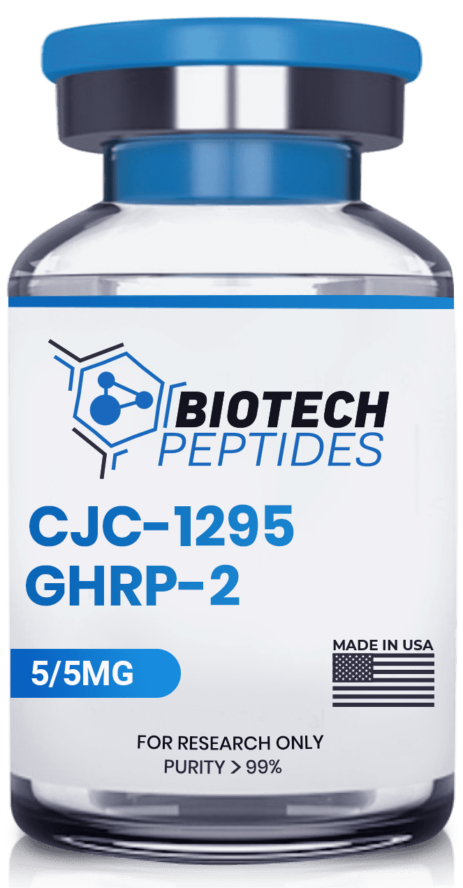
Scientific investigations have suggested that the blend of GHRP-2 and CJC-1295 peptides may be associated with the activation of discrete receptors, fostering the potential augmentation of growth hormone (GH) discharge from the anterior pituitary.

CJC-1295 represents a molecular entity that appears to exhibit a binding affinity for the growth hormone-releasing hormone (GHRH) receptors, thereby potentially instigating the secretion of growth hormone by pituitary cells. Originating from GHRH 1-29, CJC-1295 encompasses the functional sequence composed of the initial 29 amino acids of GHRH. This peptide has been subject to tetra substitution and modification via the integration of a drug affinity complex (DAC) element. The DAC component binds to plasma proteins, apparently augmenting the pharmacokinetic properties of CJC-1295.[1]

Growth Hormone Releasing Peptide 2 (GHRP-2) represents a synthetic hexapeptide comprising of six amino acids. Studies suggest that this peptide binds to the ghrelin or growth hormone secretagogues 1a receptors (GHS-R1a), which are present within the hypothalamus and the pituitary gland. Consequently, GHRP-2 appears to stimulate the production of growth hormones in pituitary cells expressing the GHS-R1a receptor.[2]

By activating distinct receptors via separate biochemical pathways, these peptides are hypothesized to synergistically elevate GH release from the anterior pituitary, potentially surpassing the individual capacities of each peptide.


GHRP-2 and CJC 1295 Peptide Blend

Both GHRP-2 and CJC-1295 have undergone extensive evaluation in diverse animal models, and are speculated to yield favorable outcomes in terms of muscle hypertrophy and hyperplasia, cardiac function, and modulation of the immune system.

Furthermore, both peptides appear to exhibit the potential to reduce blood sugar levels while promoting the accrual of lean body mass at the expense of fat deposition. Notably, these peptides are recognized for their capacity to possibly induce fat loss through the amplification of metabolic processes. 

This probable reduction in adipose tissue has been correlated with enhanced insulin sensitivity and notable advancements in cases of type 2 diabetes.


Research and Scientific Studies


Effect of GHRP-2 and CJC-1295 Blend on Growth Hormone Secretion

Scientific investigations suggest that GHRP-2 peptide appears to display considerable affinity towards the growth hormone secretagogues 1a receptor (GHS-R1a) located within the hypothalamus and the pituitary gland. In contrast, CJC-1295 appears to engage with the growth hormone-releasing hormone receptor (GHRH-R), specifically in the pituitary gland. 

Research suggests that this amalgamated peptide blend may elevate plasma growth hormone (GH) levels, implying their potential to augment GH release. Moreover, sustained release dynamics of this blend appear to prolong the half-life of the peptides, possibly leading to the extension of GH secretion compared to the individual administration of peptides. 

Studies have reported “basal GH levels may increase by 7.5-fold” contributing to an “overall 46% increase in GH secretion.”[3]

This phenomenon seems to regulate the pulsatile release of growth hormones, subject to negative feedback mechanisms, thereby potentially averting the development of supratherapeutic GH levels and their associated complications.[4]


Effect of GHRP-2 and CJC-1295 Blend on Fat Metabolism

Through distinct mechanisms, GHRP-2 and CJC-1295 peptides appear to stimulate GH release from pituitary cells, thereby facilitating fat loss. Scientific exploration indicates that CJC-1295 may exhibit a prolonged half-life compared to endogenous GHRH. 

Growth hormone is considered to demonstrate anti-obesity effects through various mechanisms, including the promotion of lipolysis, enhanced utilization of fatty acids as an energy source, and heightened fat oxidation. The peptide blend, through growth hormone release, has reportedly exhibited increased glucose synthesis and decreased glucose uptake, resulting in heightened fat utilization and storage. Studies suggest that ghrelin-like peptides such as GHRP-2 may serve as a crucial hormonal signal of nutritional status to the somatotropic axis, playing a pivotal role in integrating energy balance with the growth process.[5]


Effect of GHRP-2 and CJC-1295 Blend on Osteoporosis

A clinical study conducted on eighty postmenopausal research models of osteoporosis undergoing estrogen therapy reported significant findings regarding the effects of growth hormone (GH) peptide (such as GHRP-2 and CJC 1295) on bone health. 

The research subjects were randomized into groups receiving one of two concentrates of growth hormone or a placebo for 18 months in a double-blinded setup. The results indicated that the total body bone mineral content was notably higher in the higher concentration GH group at the 18-month mark. Over the course of 3 years, the experimental GH groups exhibited increases in total body and femoral neck bone mineral content. At the 4-year follow-up, the higher concentration GH group demonstrated a remarkable 14% increase in lumbar spine bone mineral content compared to the placebo. 

Although no disparities in bone mineral density or content were observed between groups at the 5-year follow-up, the study suggests “GH (peptides) could be used as an anabolic agent in osteoporosis[6], especially when combined with hormone replacement therapy and calcium/vitamin D supplementation.


Effect of GHRP-2 and CJC-1295 Blend on Immune System

Recent research underscores the intricate interplay between the neuroendocrine system and immune functions, illuminating a complex bidirectional relationship. 

Notably, multiple lymphoid organs, including the thymus, spleen, and peripheral blood, have been identified as sites of growth hormone (GH) production, with the GH receptor expressed across various subpopulations of lymphocytes. 

In vitro and animal studies indicate the possible role of GH in immunoregulation. GH peptides appear to stimulate the proliferation of T and B cells, facilitating immunoglobulin synthesis and promoting the maturation of myeloid progenitor cells. Moreover, GH also appears to modulate cytokine responses, thereby possibly influencing the intricate network of immune signaling.[7]

Interestingly, while GH deficiency (GHD) typically does not manifest as immunodeficiency, minor aberrations in immune function have been documented, albeit significantly less pronounced than those observed in animal models of GHD. This observation suggests the plausibility of local GH production within the immune system compensating for the absence of systemic GH. 


Disclaimer: The products mentioned are not intended for human or animal consumption. Research chemicals are intended solely for laboratory experimentation and/or in-vitro testing.  Bodily introduction of any sort is strictly prohibited by law.  All purchases are limited to licensed researchers and/or qualified professionals. All information shared in this article is for educational purposes only.


References:

  1. Ionescu M, Frohman LA. Pulsatile secretion of growth hormone (GH) persists during continuous stimulation by CJC-1295, a long-acting GH-releasing hormone analog. J Clin Endocrinol Metab. 2006 Dec;91(12):4792-7. doi: 10.1210/jc.2006-1702. Epub 2006 Oct 3. PMID: 17018654. https://pubmed.ncbi.nlm.nih.gov/17018654/ 
  2. Yamamoto D, Ikeshita N, Matsubara T, Tasaki H, Herningtyas EH, Toda K, Iida K, Takahashi Y, Kaji H, Chihara K, Okimura Y. GHRP-2, a GHS-R agonist, directly acts on myocytes to attenuate the dexamethasone-induced expressions of muscle-specific ubiquitin ligases, Atrogin-1 and MuRF1. Life Sci. 2008 Feb 27;82(9-10):460-6. doi: 10.1016/j.lfs.2007.11.019. Epub 2007 Dec 5. PMID: 18191156. https://pubmed.ncbi.nlm.nih.gov/18191156/ 
  3. Ionescu M, Frohman LA. Pulsatile secretion of growth hormone (GH) persists during continuous stimulation by CJC-1295, a long-acting GH-releasing hormone analog. J Clin Endocrinol Metab. 2006 Dec;91(12):4792-7. doi: 10.1210/jc.2006-1702. Epub 2006 Oct 3. PMID: 17018654. https://pubmed.ncbi.nlm.nih.gov/17018654/ 
  4. Sigalos JT, Pastuszak AW. The Safety and Efficacy of Growth Hormone Secretagogues. Sex Med Rev. 2018 Jan;6(1):45-53. doi: 10.1016/j.sxmr.2017.02.004. Epub 2017 Apr 8. PMID: 28400207; PMCID: PMC5632578. https://www.ncbi.nlm.nih.gov/pmc/articles/PMC5632578/ 
  5. Laferrère B, Hart AB, Bowers CY. Obese subjects respond to the stimulatory effect of the ghrelin agonist growth hormone-releasing peptide-2 on food intake. Obesity (Silver Spring). 2006 Jun;14(6):1056-63. doi: 10.1038/oby.2006.121. PMID: 16861611; PMCID: PMC2824649. https://www.ncbi.nlm.nih.gov/pmc/articles/PMC2824649/
  6. Landin-Wilhelmsen K, Nilsson A, Bosaeus I, Bengtsson BA. Growth hormone increases bone mineral content in postmenopausal osteoporosis: a randomized placebo-controlled trial. J Bone Miner Res. 2003 Mar;18(3):393-405. doi: 10.1359/jbmr.2003.18.3.393. PMID: 12619921. https://pubmed.ncbi.nlm.nih.gov/12619921/ 
  7. Meazza C, Pagani S, Travaglino P, Bozzola M. Effect of growth hormone (GH) on the immune system. Pediatr Endocrinol Rev. 2004 Aug;1 Suppl 3:490-5. PMID: 16444180. https://pubmed.ncbi.nlm.nih.gov/16444180/
Tesamorelin & Ipamorelin Blend: Studying their Potential in Growth Hormone Production

Tesamorelin & Ipamorelin Blend: Studying their Potential in Growth Hormone Production

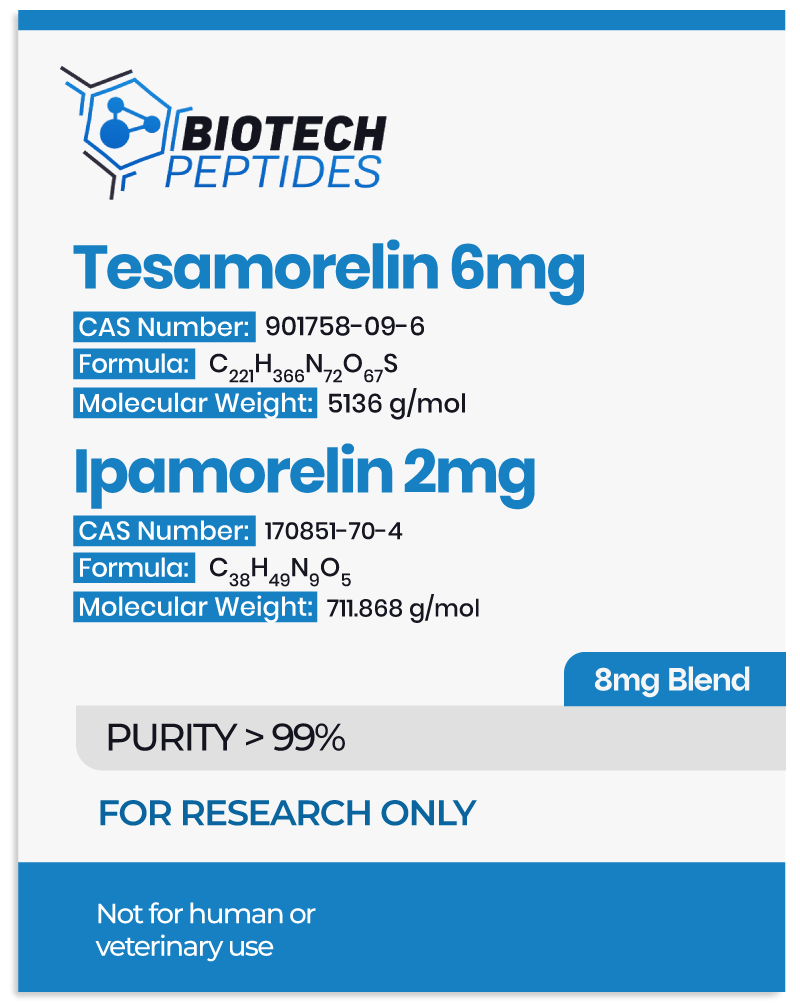
Tesamorelin and Ipamorelin represent two distinct synthetic growth hormone-releasing peptides (GHRPs) that have garnered considerable attention in the realm of biochemical research. Both compounds, Tesamorelin and Ipamorelin, appear to possess the potential to stimulate the release of endogenous growth hormone (GH) through the activation of the ghrelin receptor. This unique action has spurred increasing scientific interest in these peptides, leading to numerous investigations aimed at unraveling their molecular mechanisms and potential impacts.

Research into Tesamorelin has principally centered on its possible utility within the context of metabolic disorders, particularly in the context of HIV-associated lipodystrophy.[1] On the other hand, Ipamorelin appears to have exhibited promise in a wider range of research fields, including obesity and growth promotion.[2]

This introductory exposition explores the scientific foundation underlying Tesamorelin and Ipamorelin, and underscores their potential synergy, highlighting the importance of further exploratory research in elucidating their combined pharmacological actions.


Tesamorelin

Tesamorelin is a synthetic peptide with a specific amino acid sequence, also known as a growth hormone-releasing peptide (GHRP). Its structure consists of a chain of 44 amino acids, and it appears to function by stimulating the release of growth hormone (GH) from the pituitary gland.[3]

Tesamorelin has been studied for its potential action in research concerning conditions like HIV-associated lipodystrophy and age-related decline in GH levels. Its speculated ability to enhance GH secretion makes it an intriguing subject of research within the context of various health-related issues, although further studies are needed to fully determine its potential.


Ipamorelin

Ipamorelin is a synthetic peptide belonging to the family of growth hormone-releasing peptides (GHRPs). Its chemical structure consists of five amino acids: alanine, glutamine, histidine, leucine, and arginine.[4]

Scientists suggest that this specific sequence appears to impart Ipamorelin with the ability to stimulate the release of growth hormone (GH) by activating the ghrelin receptor, a property that potentially makes it a potent GH secretagogue. Unlike some other GHRPs, Ipamorelin appears to exhibit selective action with minimal impact on other hormones, making it an attractive candidate for research related to GH regulation.


Mechanism of Action


Tesamorelin

Tesamorelin appears to exert physiological action through the activation of growth hormone releasing hormone (GHRH) receptors localized within the anterior pituitary gland. This activation, in turn, is said to elicit an augmented synthesis and release of growth hormone (GH) into the bloodstream. Subsequently, GH appears to exert its multifaceted actions on various cell types, including hepatocytes, thereby prompting the synthesis of insulin-like growth factor-1 (IGF-1). IGF-1, akin to GH, is considered to play a pivotal role in cellular growth promotion, apoptosis inhibition, glucose regulation, and lipolysis.[5]

Notably, study findings have indicated that research models of HIV-associated lipodystrophy often manifest diminished levels of GH and IGF-1. Tesamorelin may rectify this imbalance, possibly managing overall lipid metabolism and accumulation. 

An intriguing aspect of Tesamorelin lies in its structural modification at the N-terminus, diverging from natural GHRH. This structural alteration appears to enhance peptide stability and may confer heightened resistance to enzymatic deactivation.


Ipamorelin

Ipamorelin represents a pioneering synthetic growth hormone secretagogue that appears to operate by enhancing the endogenous production of growth hormone. It is speculated to elicit the release of growth hormone from the anterior pituitary gland, thereby sustaining physiological hormone levels. 

This peptide appears to exhibit a remarkable degree of specificity for the growth hormone-releasing peptide (GHRP) receptors situated within the pituitary gland. Analogous to other GHRP receptor agonists, Ipamorelin may engage these receptors to stimulate the secretion of growth hormone, hGH.[6]


Research and Scientific Studies


Tesamorelin and Ipamorelin Peptide Blend and Growth Hormone Deficiency

The Tesamorelin and Ipamorelin blend appears to exhibit a multifaceted mechanism of action that, when combined, may offer synergistic effects in the context of growth hormone deficiency. 

When introduced concurrently, the Tesamorelin and Ipamorelin blend has been suggested by researchers to manifest a dual-action approach. Tesamorelin appears to stimulate GH release via GHRH receptor activation, while Ipamorelin seems to directly stimulate GHSR, apparently leading to increased GH secretion. This combined action is presumed to amplify the overall GH response, potentially offering advantages in terms of optimizing mass composition, lipid profiles, insulin sensitivity, and overall metabolic functionality. 

By addressing different components of the growth hormone axis through their distinct mechanisms, Tesamorelin and Ipamorelin may possibly enhance, collectively, pituitary gland activity that ultimately facilitates the release of endogenous growth hormone.[7]


Tesamorelin and Ipamorelin Peptide Blend and Lipodystrophy

Lipodystrophy often accompanies insulin resistance, dyslipidemia, and an elevated susceptibility to cardiovascular complications. Clinical research has indicated that the introduction of Tesamorelin to lipodystrophic research subjects may engender a noteworthy reduction in visceral adipose tissue (VAT) alongside possible enhancements in insulin sensitivity and lipid profiles. 

Tesamorelin’s mechanism of action appears to involve the activation of the growth hormone-releasing hormone receptor (GHRHR), potentially fostering the secretion of endogenous growth hormone and facilitating lipolysis. As a result, Tesamorelin may contribute to the preservation of abdominal subcutaneous adipose tissue and favorable alterations in lipid metabolism.

In parallel, Ipamorelin has exhibited signs of promise in modulating adipose tissue metabolism. The conjecture surrounding the combined utilization of Tesamorelin and Ipamorelin postulates synergistic effects, which may further augment the reduction of VAT and ameliorate metabolic parameters in models of lipodystrophy.[8]


Tesamorelin and Ipamorelin Peptide Blend and Cognitive Improvement

Growing study data suggests that growth hormone and its secretagogues, including Tesamorelin and Ipamorelin, may exert modulatory influences on neuroplasticity, neuronal survival, and synaptic plasticity, all pivotal elements for optimal cognitive function. Preclinical investigations have revealed that Tesamorelin may potentially enhance memory and learning capacities, possibly via neurogenesis and synaptic plasticity. 

Concurrently, Ipamorelin appears to have some potential to augment spatial memory and cognitive function in animal models. The combined presentation of Tesamorelin and Ipamorelin may prospectively magnify these cognitive benefits through their proposed synergistic mechanisms of action. However, the validation of these findings and the elucidation of the blend’s biological effects on cognitive functioning necessitate future study.


Tesamorelin and Ipamorelin Peptide Blend and Type 2 Diabetes

The combination of Tesamorelin and Ipamorelin has garnered attention of researchers within the context of Type 2 Diabetes Mellitus (T2DM) due to promising outcomes observed in exploratory investigations. Both peptides appear to exhibit potential for enhancing glycemic control and ameliorating the metabolic irregularities associated with T2DM. 

Tesamorelin’s mechanism of action is speculated to involve the stimulation of endogenous growth hormone secretion, a process linked to improved insulin sensitivity and enhanced glucose utilization. Ipamorelin similarly has been suggested to influence glucose metabolism and insulin sensitivity. When presented together, Tesamorelin and Ipamorelin may confer complementary impacts, potentially encompassing reductions in hemoglobin A1c (HbA1c) levels, enhancements in insulin sensitivity, and reductions in visceral adiposity in the case of T2DM.[9]


Tesamorelin and Ipamorelin Peptide Blend and Pituitary Glad

The combination of Tesamorelin and Ipamorelin may have a notable influence on the pituitary gland, a pivotal endocrine organ responsible for the regulation of growth hormone secretion. 

Research indicates that Tesamorelin may possess a specific affinity for the growth hormone-releasing hormone receptor (GHRHR), potentially initiating the intracellular signaling cascade leading to the synthesis and subsequent release of growth hormone. As supported by previous studies, “Tesamorelin, as a synthetic growth hormone-releasing hormone, exerts its action on the anterior pituitary gland to stimulate endogenous growth hormone secretion”[10]

In contrast, Ipamorelin exhibits potent agonistic activity toward the growth hormone secretagogue receptor (GHSR), possibly facilitating the secretion of growth hormone. As documented in scientific literature, “Ipamorelin stands as the pioneering GHRP-receptor agonist, demonstrating a selectivity for GH release akin to that of GHRH. The specificity inherent in Ipamorelin positions it as a compelling candidate for prospective clinical development”[11]

When introduced together, these peptides appear to elicit a synergistic effect on the pituitary gland, potentially resulting in heightened production of growth hormone. This cooperative interplay between Tesamorelin and Ipamorelin holds promise as a valuable avenue for the optimization of growth hormone levels.[7]


In Summary

The combination of Tesamorelin and Ipamorelin peptides, when introduced concurrently, presents an intriguing prospect for magnifying biological benefits through their proposed complementary modes of action. By jointly stimulating the growth hormone axis and modulating neurotrophic factors, this peptide blend may hold promise as a catalyst for cognitive improvement. 

The majority of these findings stem from research conducted in animal models. Therefore, ongoing research remains imperative to fully harness the benefits of these peptides and explore their relevance in the realm of cognitive improvement across a wider variety of species.

Disclaimer: The products mentioned are not intended for human or animal consumption. Research chemicals are intended solely for laboratory experimentation and/or in-vitro testing.  Bodily introduction of any sort is strictly prohibited by law.  All purchases are limited to licensed researchers and/or qualified professionals. All information shared in this article is for educational purposes only.


References:

  1. Dhillon S. Tesamorelin: a review of its use in the management of HIV-associated lipodystrophy. Drugs. 2011 May 28;71(8):1071-91. doi: 10.2165/11202240-000000000-00000. PMID: 21668043. https://pubmed.ncbi.nlm.nih.gov/21668043/ 
  2. Gao Y, Yuan X, Zhu Z, Wang D, Liu Q, Gu W. Research and prospect of peptides for use in obesity treatment (Review). Exp Ther Med. 2020 Dec;20(6):234. doi: 10.3892/etm.2020.9364. Epub 2020 Oct 16. PMID: 33149788; PMCID: PMC7604735. https://www.ncbi.nlm.nih.gov/pmc/articles/PMC7604735/ 
  3. National Center for Biotechnology Information (2023). PubChem Compound Summary for , Tesamorelin. https://pubchem.ncbi.nlm.nih.gov/compound/Tesamorelin
  4. National Center for Biotechnology Information (2023). PubChem Compound Summary for CID 9831659, Ipamorelin. https://pubchem.ncbi.nlm.nih.gov/compound/Ipamorelin
  5. LiverTox: Clinical and Research Information on Drug-Induced Liver Injury [Internet]. Bethesda (MD): National Institute of Diabetes and Digestive and Kidney Diseases; 2012-. Tesamorelin. [Updated 2018 Oct 20]. Available from: https://www.ncbi.nlm.nih.gov/books/NBK548730/ 
  6. K. Raun et al., Ipamorelin, the first selective growth hormone secretagogue, European Journal of Endocrinology, November 1998, 139 552-561.  https://pubmed.ncbi.nlm.nih.gov/9849822/ 
  7. Rogério G. Gondo et al, Growth Hormone-Releasing Peptide-2 Stimulates GH Secretion in GH-Deficient Patients with Mutated GH-Releasing Hormone Receptor, The Journal of Clinical Endocrinology & Metabolism, Volume 86, Issue 7, 1 July 2001, Pages 3279–3283, https://doi.org/10.1210/jcem.86.7.7694 
  8. Falutz J, Mamputu JC, Potvin D, Moyle G, Soulban G, Loughrey H, Marsolais C, Turner R, Grinspoon S. Effects of tesamorelin (TH9507), a growth hormone-releasing factor analog, in human immunodeficiency virus-infected patients with excess abdominal fat: a pooled analysis of two multicenter, double-blind placebo-controlled phase 3 trials with safety extension data. J Clin Endocrinol Metab. 2010 Sep;95(9):4291-304. doi: 10.1210/jc.2010-0490. Epub 2010 Jun 16. PMID: 20554713. https://pubmed.ncbi.nlm.nih.gov/20554713 
  9. Clemmons DR, Miller S, Mamputu JC. Safety and metabolic effects of tesamorelin, a growth hormone-releasing factor analogue, in patients with type 2 diabetes: A randomized, placebo-controlled trial. PLoS One. 2017 Jun 15;12(6):e0179538. doi: 10.1371/journal.pone.0179538. PMID: 28617838; PMCID: PMC5472315. https://www.ncbi.nlm.nih.gov/pmc/articles/PMC5472315/  
  10. Adrian S, Scherzinger A, Sanyal A, Lake JE, Falutz J, Dubé MP, Stanley T, Grinspoon S, Mamputu JC, Marsolais C, Brown TT, Erlandson KM. The Growth Hormone Releasing Hormone Analogue, Tesamorelin, Decreases Muscle Fat and Increases Muscle Area in Adults with HIV. J Frailty Aging. 2019;8(3):154-159. doi: 10.14283/jfa.2018.45. PMID: 31237318; PMCID: PMC6766405. https://www.ncbi.nlm.nih.gov/pmc/articles/PMC6766405/ 
  11. Raun K, Hansen BS, Johansen NL, Thøgersen H, Madsen K, Ankersen M, Andersen PH. Ipamorelin, the first selective growth hormone secretagogue. Eur J Endocrinol. 1998 Nov;139(5):552-61. doi: 10.1530/eje.0.1390552. PMID: 9849822. https://pubmed.ncbi.nlm.nih.gov/9849822/