PNC-27 Research

PNC-27 Research

PNC-27 peptide is a synthetic compound created for implications in cancer cell research. It is a peptide from the PNC family of investigational proteins designed to target cancerous cells, induce cell necrosis, and leave healthy, normal cells unharmed and intact.

PNC-27 consists of an HDM-2 binding domain similar to p53 residues 12-26 and a transmembrane-penetrating portion. PNC-27 peptide has been suggested by researchers to bind to and kills cancerous cells via membrane lysis, or the breakdown of the cell membrane, according to study findings.


PNC-27 Peptide Overview

Dr. Ehsan Sarafraz-Yazdi and other researchers have suggested that PNC-27 may impact cancer cells, in a 2010 research article published by the American Association for Cancer.

According to the study’s authors, the mechanism of action of PNC-27 appears to result from oligomeric pores developing in the plasma membrane of tumor cells. Untransformed or non-tumor cells do not appear to form oligomeric pores. PNC-27 perceived selectivity on cancer cells may be caused by MDM2’s mis-localization to cancerous cell plasma membranes.

These hypotheses are believed by scientists to describe the distinct localization of MDM2 variants in cancerous cell plasma membranes.


PNC-27 Peptide Action

PNC-27 was initially developed for employment in HIV research, but its anti-cancer potential, which included killing cancer cells while leaving normal cells alone, emerged and thus changed its initial research focus.

The PNC-27 peptide does not appear to impact cells other than cancerous cells. PNC-27 has been suggested to bind to individual cancer cell membranes, causing holes to form. Due to the difference in osmotic pressure between the inside and outside of the tumor cells, the holes may cause a rapid implosion, resulting in immediate cell death.

HDM-2 is considered to be found in the cell membranes of cancer cells. When PNC-27 is introduced, the peptide may immediately move to bind to the HDM-2 peptide in the plasma membrane of cancerous cells. PNC-27 may induce damage by inducing membrane lysis and, thus, cancer cell death by binding to the HDM-2 peptides.


PNC-27 Research Studies

According to PNC-27 research in animal research models, the peptide may reduce pain perception on a short term basis. The animal models developed flu-like consequences during the third week of exposure, indicating that the animal’s immune system may have reacted to the death of cancerous cells.

Bilirubin and lactate dehydrogenase appear to levels rise during the sixth week of PNC-27 exposure. Around the tenth week, the tumors may potentially soften and become more pliable, resulting in significant tumor breakdown.

The tumor’s size may grow more extensive in the tenth week. In any case, it may be due to inflammation due to the immune system’s response. Animal research models’ energy levels reportedly increased, and cancer-related symptoms decreased in the third month.

The results of the PNC-27 studies suggested that the peptide may potentially impact the cell membranes of cancerous cells. Furthermore, the noncancerous or non-transformed cells surrounding the cancerous cells appeared to remain unimpacted. Moreover, research results suggest that PNC-27 may influence specific markers in cancerous cell membranes and that PNC-27 may exhibit a longer half-life via this mechanism of action.


Examining PNC-27 in Relation to Cancer Cells

According to research, the peptide may potentially inhibit pancreatic cancer cell growth. In different tumor cell lines, including BMRPA—ras transformed rat acinar pancreatic carcinoma cell line, PNC-27 induces necrosis but not apoptosis. Notably, PNC-27 was not suggested to affect noncancerous cells. According to scientific findings, the peptide appeared to induce a complete blockade of tumor growth during the second week of and after exposure, resulting in weak growth of tumor cells that plateaued at small tumor sizes compared to tumor growth in the absence of the peptide. PNC-27 exposure after the appearance of tumor growth at a distant site reportedly resulted in a decrease in tumor size followed by a slow increase in tumor growth.

According to studies, PNC-27 may potentially induce tumor cell necrosis in a poorly developed non-solid leukemia cell line based on HDM-2 expression in the cells’ plasma membrane. PNC-27 peptide may kill solid tumor cells by binding to HDM-2 proteins within their cell membranes, bypassing the p53 pathway. PNC-27 peptide might also induce necrosis via membrane lysis in solid tissue tumor cells by binding to HDM-2, independent of the p53 pathway.


Disclaimer: The products mentioned are not intended for human or animal consumption. Research chemicals are intended solely for laboratory experimentation and/or in-vitro testing.  Bodily introduction of any sort is strictly prohibited by law.  All purchases are limited to licensed researchers and/or qualified professionals. All information shared in this article is for educational purposes only.


References


  1. Sarafraz-Yazdi E, Bowne WB, Adler V, Sookraj KA, Wu V, Shteyler V, Patel H, Oxbury W, Brandt-Rauf P, Zenilman ME, Michl J, Pincus MR. Anticancer peptide PNC-27 adopts an HDM-2-binding conformation and kills cancer cells by binding to HDM-2 in their membranes. Proc Natl Acad Sci U S A. 2010 Feb 2;107(5):1918-23. doi: 10.1073/pnas.0909364107. Epub 2010 Jan 11. PMID: 20080680; PMCID: PMC2836618.
  2. Sarafraz-Yazdi E, Mumin S, Cheung D, Fridman D, Lin B, Wong L, Rosal R, Rudolph R, Frenkel M, Thadi A, Morano WF, Bowne WB, Pincus MR, Michl J. PNC-27, a Chimeric p53-Penetratin Peptide Binds to HDM-2 in a p53 Peptide-like Structure, Induces Selective Membrane-Pore Formation and Leads to Cancer Cell Lysis. Biomedicines. 2022 Apr 20;10(5):945. doi: 10.3390/biomedicines10050945. PMID: 35625682; PMCID: PMC9138867.
  3. Davitt K, Babcock BD, Fenelus M, Poon CK, Sarkar A, Trivigno V, Zolkind PA, Matthew SM, Grin’kina N, Orynbayeva Z, Shaikh MF, Adler V, Michl J, Sarafraz-Yazdi E, Pincus MR, Bowne WB. The anti-cancer peptide, PNC-27, induces tumor cell necrosis of a poorly differentiated non-solid tissue human leukemia cell line that depends on expression of HDM-2 in the plasma membrane of these cells. Ann Clin Lab Sci. 2014 Summer;44(3):241-8. PMID: 25117093.
  4. Alagkiozidis I, Gorelick C, Shah T, Chen YA, Gupta V, Stefanov D, Amarnani A, Lee YC, Abulafia O, Sarafraz-Yazdi E, Michl J. Synergy between Paclitaxel and Anti-Cancer Peptide PNC-27 in the Treatment of Ovarian Cancer. Ann Clin Lab Sci. 2017 May;47(3):271-281. PMID: 28667027.
  5. Thadi A, Lewis L, Goldstein E, Aggarwal A, Khalili M, Steele L, Polyak B, Seydafkan S, Bluth MH, Ward KA, Styler M, Campbell PM, Pincus MR, Bowne WB. Targeting Membrane HDM-2 by PNC-27 Induces Necrosis in Leukemia Cells But Not in Normal Hematopoietic Cells. Anticancer Res. 2020 Sep;40(9):4857-4867. doi: 10.21873/anticanres.14488. PMID: 32878773.
The Potential Function of Kisspeptin-10

The Potential Function of Kisspeptin-10

According to research and studies, Kisspeptin is a naturally occurring protein known as metastin that may play a vital role in reproductive hormone secretion. Kisspeptin-10 peptide is suggested to regulate testosterone levels and reproductive behaviors. The peptide has also been hypothesized to influence hormone signaling which may influence mood and behavior, and has also been implicated in the regulation of kidney function, and in angiogenesis. Kisspeptin-10 peptide may also potentially inhibit tumor growth and metastasis in the brain. The peptide is under study for its potential to modulate gonadotropin-releasing hormone (GnRH).[1]
 

Testosterone Secretion

Kisspeptin-10 peptide may affect and influence testosterone levels by modulating FSH and LH levels in the circulatory system, which is sex-specific. Kisspeptin-10 might increase testosterone levels in male species but has no perceived effect on testosterone levels in female species.

Kisspeptin-10 peptide analogs may influence the pulse frequency of LH in males, implying that Kisspeptin-10 might possibly fine-tune the regular pulsatile release of sex hormones.

Studies examining Kisspeptin-10 in male organisms suggest a concentration-dependent and rapid increase in serum LH and testosterone levels. Kisspeptin-10 may accomplish this by increasing the pulsatile release of LH. High concentrations of Kisspeptin-10 peptide may cause rapid pulsation, resulting in continuous LH release.


Gonadotropin-Releasing Hormone

GnRH neurons in the hypothalamus appear to synthesize and release gonadotropin-releasing hormone (GnRH). The first hypothalamic-pituitary-gonadal hormonal release may influence the anterior pituitary gland to release FSH and luteinizing hormone (LH). The primary hormone for reproductive development and the control of gamete maturation in the genitals is considered to be GnRH.


Kisspeptin-10 Peptide and Energy

Kisspeptin-10 peptide neurons may be energy status sensitive: undernutrition and overnutrition may inhibit Kisspeptin-10 neurons’ ability to induce GnRH release.[3] Infertility may occur in males and females alike under extreme energy balance conditions, mediated by the Kisspeptin-10 peptide. As a result, Kisspeptin-10 peptide synthesis and release may be energy balance sensitive. However, the findings suggest that Kisspeptin-10 may be the sole regulator of energy balance.

Studies in mice samples depleted of the Kisspeptin-10 receptor (Kiss1r) via genetic manipulation suggest an increase in adiposity and a decrease in energy expenditure. Studies suggest that Kisspeptin-10 receptors exist in adipose (fat) and brown adipose tissues. Kisspeptin-10 peptide may also help to explain the neurochemical modulation that results in energy-modulating behavior concerning reproduction.


Kisspeptin-10 Peptide and Mood

A study examining Kisspeptin suggested that the peptide may have increased limbic brain activity.[4] There was a reported increase in drive, reward-seeking behavior, and overall improved mood. Kisspeptin-10 peptide appeared to accomplish this by integrating the emotional brain and the sexual process through the overall reproduction process.


Kisspeptin-10 Peptide and the Heart

Kisspeptin-10 peptide and its receptors are considered to be found in numerous locations throughout the kidney and may play an essential role in stimulating kidney functions, according to the findings. Animal studies in mice lacking the Kiss1 receptor suggest that the peptide may be critical for maintaining glomerular development during development, though whether this action is direct or indirect is unknown.

Although the function of Kisspeptin-10 peptide in the kidney is unknown, the peptide may influence vascular development and injury responses. Kisspeptin-10 studies in mice with cardiovascular disease suggest that the peptide may act in the vascular beds, controlling vasoconstriction.

The potential of Kisspeptin-10 peptide on angiogenesis and vascular function may significantly impact the kidney and cardiovascular system.


Kisspeptin-10 Peptide and Memory

Research studies in mice suggest that Kisspeptin analogs may reverse the navigational and learning impairment caused by ethanol intoxication. Kisspeptin-10 peptide and its analogs may potentially play a role in neurons encoding information and compensating for learning defects in certain genetic and chronic disease conditions.


Kisspeptin-10 Peptide and Cancer

Kisspeptin-10 peptide was discovered about 20 years ago and suggested to suppress melanoma metastasis by approximately 95%, indicating that the peptide may have an effect to reduce cancer cell spread. The evaluation of various metastatic cancers suggests that Kisspeptin-10 levels may decrease in the bladder, GI, breast, pancreatic, ovarian, thyroid, skin, and prostate, suggesting that the peptide may exert some role in cancer spread.

In 2020, an experiment on Kisspeptin-10 peptide levels under daylight exposure established a potential link between melatonin, kisspeptin-10, and cancer.[5] Findings in mice models exposed to daylight supported findings of high levels of Kisspeptin and low melatonin levels, and vice versa in the dark. In daylight mice, there was no appearance of increase in tumor growth rates, metastasis, or volume. Kisspeptin-10 and melatonin may be implicated in tumor suppression as one appears to influence the actions of the other, though the precise relationship is unknown.


Disclaimer: The products mentioned are not intended for human or animal consumption. Research chemicals are intended solely for laboratory experimentation and/or in-vitro testing.  Bodily introduction of any sort is strictly prohibited by law.  All purchases are limited to licensed researchers and/or qualified professionals. All information shared in this article is for educational purposes only.


References


  1. Jayasena CN, Nijher GM, Comninos AN, Abbara A, Januszewki A, Vaal ML, Sriskandarajah L, Murphy KG, Farzad Z, Ghatei MA, Bloom SR, Dhillo WS. The effects of kisspeptin-10 on reproductive hormone release show sexual dimorphism in humans. J Clin Endocrinol Metab. 2011 Dec;96(12):E1963-72. doi: 10.1210/jc.2011-1408. Epub 2011 Oct 5. PMID: 21976724; PMCID: PMC3232613.
  2. George JT, Veldhuis JD, Roseweir AK, Newton CL, Faccenda E, Millar RP, Anderson RA. Kisspeptin-10 is a potent stimulator of LH and increases pulse frequency in men. J Clin Endocrinol Metab. 2011 Aug;96(8):E1228-36. doi: 10.1210/jc.2011-0089. Epub 2011 Jun 1. PMID: 21632807; PMCID: PMC3380939.
  3. Navarro VM. Metabolic regulation of kisspeptin – the link between energy balance and reproduction. Nat Rev Endocrinol. 2020 Aug;16(8):407-420. doi: 10.1038/s41574-020-0363-7. Epub 2020 May 19. PMID: 32427949; PMCID: PMC8852368.
  4. Comninos AN, Wall MB, Demetriou L, Shah AJ, Clarke SA, Narayanaswamy S, Nesbitt A, Izzi-Engbeaya C, Prague JK, Abbara A, Ratnasabapathy R, Salem V, Nijher GM, Jayasena CN, Tanner M, Bassett P, Mehta A, Rabiner EA, Hönigsperger C, Silva MR, Brandtzaeg OK, Lundanes E, Wilson SR, Brown RC, Thomas SA, Bloom SR, Dhillo WS. Kisspeptin modulates sexual and emotional brain processing in humans. J Clin Invest. 2017 Feb 1;127(2):709-719. doi: 10.1172/JCI89519. Epub 2017 Jan 23. PMID: 28112678; PMCID: PMC5272173.
  5. Pazarci P, Kaplan H, Alptekin D, Yilmaz M, Lüleyap U, Singirik E, Pelit A, Kasap H, Yegani A. The effects of daylight exposure on melatonin levels, Kiss1 expression, and melanoma formation in mice. Croat Med J. 2020 Feb 29;61(1):55-61. doi: 10.3325/cmj.2020.61.55. PMID: 32118379; PMCID: PMC7063558.
Selank Studies in Anxiety and Brain Function

Selank Studies in Anxiety and Brain Function

Selank is a short peptide with anxiolytic and nootropic potential, structurally similar to the naturally occurring Tuftsin. Tuftsin is a tetrapeptide that appears to stimulate the immune system. T helper cells, monoamine neurotransmitters, IL-6, and brain-derived neurotrophic factors all appear to be regulated by it (BDNF). Selank peptide only differs from Tuftsin by an extra four amino acids in its chain that researchers suggest may induce a longer half-life and improve stability.
 

Selank Peptide Overview

Research suggests that Selank may exhibit certain neuroprotective characteristics. Selank peptide may support mood and potentially decrease stress hormone secretion.

According to research, 84 genes have been linked to GABA signaling.[2] Selank peptide potentially regulates 7 genes, while 45 genes may induce change in expression. The net result suggests that 52 genes tied to GABA signaling may be impacted by Selank to a certain degree, suggesting that Selank may directly modulate gene expression in nerve cells and impact changes by influencing the affinity of the GABA receptor for GABA.

The impact of Selank on enkephalin degradation may potentially regulate effects on GABA receptors. According to scientific data, research models exhibit increased enkephalinase activity in the blood after stress hormone secretion, resulting in short half-lived enkephalins.

Selank peptide potentially resets the enzymatic pathway to protect the natural anxiolytic peptides by inhibiting enkephalin degradation. The research proposes that part of the Selank action may be from a preventive impact on enkephalin degradation.


Selank Peptide and Memory

Scientists believe Selank peptide may enhance certain cognitive processes.[3] Training mice with rewards, such as food, in combination with Selank, may potentially boost memory trace stability and storage. Selank’s impact in these mice models, despite anxiety levels, indicates that the peptide goes a long way to lower stress-related memory impairment.

Selank peptide may also potentially alter memory by regulating gene expression in the hippocampus. Research suggests changes in mRNA levels of 36 varying genes. Most genes encode proteins relating to the plasma membrane, regulating ion-dependent processes in memory and learning.

According to studies, Selank may potentially salvage learning and memory recall. Exposing mice samples to a neurotoxin and Selank suggested a restoration of cognitive processes. This potential correlates with an artificial inhibition of the catecholamine system in the brain via Selank. Scientists believe that Selank peptide may offer cognitive support following traumatic brain injury.


Selank Peptide and Pain Perception

Selank peptide may potentially inhibit enzymes in the blood that induce enkephalin synthesis, reducing natural enkephalin degradation. Enkephalins are peptides that bind to opioid receptors, reducing the severity of pain. They also regulate stress response and are at abnormally high levels in the adrenal glands and brain. Therefore, Selank peptide may possibly modulate stress response and its impacts on learning, concentration, and memory by lowering enkephalin levels in the brain.


Selank and the Immune System following Anxiety

Selank peptide may potentially suppress genes that control inflammatory cytokine inflammation, IL-6, in research models of depression.[4] Selank’s research in mice models suggests that the peptide may control the expression of 34 genes that influence the inflammatory process. These genes may impact cytokines, chemokines, and both receptors. Specifically, Selank peptide modulates Bcl6 expression, a gene that controls immune system development.

In addition, Selank peptide and Selank fragments may temporarily influence gene expression for Xcr1, C3, CAsp1, and II2rf in the mouse spleen. Selank may affect the immune system balance and inflammation by impacting these genes.


Disclaimer: The products mentioned are not intended for human or animal consumption. Research chemicals are intended solely for laboratory experimentation and/or in-vitro testing.  Bodily introduction of any sort is strictly prohibited by law.  All purchases are limited to licensed researchers and/or qualified professionals. All information shared in this article is for educational purposes only.


References


  1. Medvedev VE, Tereshchenko ON, Israelian AIu, Chobanu IK, Kost NV, Sokolov OIu, Miasoedov NF. [A comparison of the anxiolytic effect and tolerability of selank and phenazepam in the treatment of anxiety disorders]. Zh Nevrol Psikhiatr Im S S Korsakova. 2014;114(7):17-22. Russian. PMID: 25176261.
  2. Volkova A, Shadrina M, Kolomin T, Andreeva L, Limborska S, Myasoedov N, Slominsky P. Selank peptide Administration Affects the Expression of Some Genes Involved in GABAergic Neurotransmission. Front Pharmacol. 2016 Feb 18;7:31. doi: 10.3389/fphar.2016.00031. PMID: 26924987; PMCID: PMC4757669.
  3. Semenova TP, Kozlovskaya MM, Zakharova NM, Kozlovskii II, Zuikov AV. Effect of selank on cognitive processes after damage inflicted to the cerebral catecholamine system during early ontogeny. Bull Exp Biol Med. 2007 Nov;144(5):689-91. doi: 10.1007/s10517-007-0406-2. PMID: 18683497.
  4. Uchakina ON, Uchakin PN, Miasoedov NF, Andreeva LA, Shcherbenko VE, Mezentseva MV, Gabaeva MV, Sokolov OIu, Zozulia AA, Ershov FI. [Immunomodulatory effects of selank in patients with anxiety-asthenic disorders]. Zh Nevrol Psikhiatr Im S S Korsakova. 2008;108(5):71-5. Russian. PMID: 18577961.
Sermorelin & Ipamorelin Blend: Potential Analogues of Growth Hormone

Sermorelin & Ipamorelin Blend: Potential Analogues of Growth Hormone


Ipamorelin & Sermorelin Blend

Researchers suggest that endogenous growth hormone production may increase under the influence of Ipamorelin and Sermorelin in animal research models. It is hypothesized that Sermorelin may exhibit different mechanisms of action in the pituitary gland than Ipamorelin. As a result, a higher growth hormone level may be obtained upon combinatorial introduction of Ipamorelin and Sermorelin.


Ipamorelin

Ipamorelin is considered by researchers to be the most direct ghrelin analog or agonist of the growth hormone secretagogue. The peptide appears to potentially increase serum growth hormone levels in certain animals.[1] Ipamorelin appears to stimulate the pituitary gland-based secretion of growth-related secretions. It is suggested to be impactful in skeletal and muscular tissue growth and repair.

Ipamorelin may potentially stimulate the production of insulin-like growth factor 1 (IGF-1), which may increase contractile force in muscle fibers. According to studies, Ipamorelin does not appear to affect cortisol, ghrelin, or prolactin levels.[3] The researchers also reported that “Ipamorelin is the first GHRP-receptor agonist with a selectivity for GH release similar to that displayed by GHRH. The specificity of ipamorelin makes this compound a very interesting candidate for future [research].”


Sermorelin

Sermorelin is a growth hormone analog that researchers suggest mimics the primary function of GHRH while avoiding the ancillary action. Sermorelin, like the natural GHRH mechanism, appears to bind to the growth hormone-releasing hormone receptor (GHRHr) in the anterior pituitary gland.[4] The peptide may increase endogenous IGF-1 secretion while potentially promoting robust growth hormone production.

A negative feedback mechanism induced by somatostatin may control the positive effect of Sermorelin on the release of the growth hormone. Somatostatin is a growth-inhibiting hormone (GHIH) that may inhibit the secretion of other hormones in the endocrine system as well as certain exocrine secretions.

Sermorelin’s natural action mechanism may preserve the hypothalamic-pituitary-somatotropic axis, preventing the cessation of direct replacement with hGH.

Sermorelin is researched for its potential to influence growth hormone secretion.[5] The researchers report that “Sermorelin stimulates pituitary gene transcription of hGH messenger RNA, increasing pituitary reserve and thereby preserving more of the growth hormone neuroendocrine axis, which is the first to fail during aging.” The peptide may improve bone density, renal function, seizure activity, the adverse action of dementia, and scarring caused by a heart attack. Sermorelin may also promote angiogenesis, reduce cardiomyocyte cell death, and reduce inflammation after cardiac injury. Sermorelin may also potentially improve sleep quality, the central nervous system, bone function, bowel motility, and diabetes insulin control.


Disclaimer: The products mentioned are not intended for human or animal consumption. Research chemicals are intended solely for laboratory experimentation and/or in-vitro testing.  Bodily introduction of any sort is strictly prohibited by law.  All purchases are limited to licensed researchers and/or qualified professionals. All information shared in this article is for educational purposes only.
 

References


  1. Johansen PB, Nowak J, Skjaerbaek C, Flyvbjerg A, Andreassen TT, Wilken M, Orskov H. Ipamorelin, a new growth-hormone-releasing peptide, induces longitudinal bone growth in rats. Growth Horm IGF Res. 1999 Apr;9(2):106-13. doi: 10.1054/ghir.1999.9998. PMID: 10373343.
  2. Johansen PB, Segev Y, Landau D, Phillip M, Flyvbjerg A. Growth hormone (GH) hypersecretion and GH receptor resistance in streptozotocin diabetic mice in response to a GH secretagogue. Exp Diabesity Res. 2003 Apr-Jun;4(2):73-81. doi: 10.1155/EDR.2003.73. PMID: 14630569; PMCID: PMC2478601.
  3. Raun K, Hansen BS, Johansen NL, Thøgersen H, Madsen K, Ankersen M, Andersen PH. Ipamorelin, the first selective growth hormone secretagogue. Eur J Endocrinol. 1998 Nov;139(5):552-61. doi: 10.1530/eje.0.1390552. PMID: 9849822.
  4. Prakash A, Goa KL. Sermorelin: a review of its use in the diagnosis and treatment of children with idiopathic growth hormone deficiency. BioDrugs. 1999 Aug;12(2):139-57. doi: 10.2165/00063030-199912020-00007. PMID: 18031173.
  5. Walker RF. Sermorelin: a better approach to management of adult-onset growth hormone insufficiency? Clin Interv Aging. 2006;1(4):307-8. doi: 10.2147/ciia.2006.1.4.307. PMID: 18046908; PMCID: PMC2699646.
  6. Chang Y, Huang R, Zhai Y, Huang L, Feng Y, Wang D, Chai R, Zhang W, Hu H. A potentially effective drug for patients with recurrent glioma: sermorelin. Ann Transl Med. 2021 Mar;9(5):406. doi: 10.21037/atm-20-6561. PMID: 33842627; PMCID: PMC8033379.
Research of AOD 9604 in Fat Reduction and Metabolic Regulation

Research of AOD 9604 in Fat Reduction and Metabolic Regulation

 
AOD9604 peptide is a synthetic version of growth hormone (HGH), a revised version of peptide fragment 176-191 with a disulfide bridge. Despite the peptide’s primary potential being hypothesized in relation to fat reduction (lipolysis), scientific data suggests that it may exert action in the context of heart disease, hypercholesterolemia (the metabolic syndrome), osteoarthritis, and cartilage restoration.

While inhibiting lipogenesis, AOD9604 may potentially stimulate and improve lipolysis (fat disintegration or breakdown). The peptide does not appear to affect IGF-1 or insulin levels and may not have any association in diabetes or glucose intolerance.[1] Researchers comment that “chronic [exposure to] AOD9604 showed no adverse effect on insulin sensitivity of the animals, as [seen] with euglycemic clamp techniques.” There is also no data to suggest that the peptide leads to the formation of antibodies. [2]


AOD9604 and General Research

The peptide appears to stimulate the pituitary gland by acting directly on it. This appears to be the same mechanism of action as GH, as it mimics the actions of the natural growth hormone on body fat metabolism. AOD9604 has been suggested by researchers to accelerate fat-burning processes and restore anabolism by acting on the pituitary gland. The compound has not been implicated in appetite alteration or blood sugar levels.

AOD9604 research in mice suggested that the peptide may impact more than the beta-3-adrenergic receptors on white fat.[4] The scientists suggested that “both hGH and AOD9604 are capable of increasing the repressed levels of beta(3)-AR RNA in obese mice to levels comparable with those in lean mice.” Initially, it was assumed that AOD9604 may increase the metabolic rate in fat cells by binding to these receptors and switching them from storage to user mode. However, even mice lacking these receptors appeared to have reduced fat cell accumulation after being exposed to AOD9604.

Because the beta-3-adrenergic receptor appears to influence fat loss through AOD9604, another mechanism may potentially be at work. The compound may indirectly stimulate apoptosis in white fat cells, according to this theory.

Finally, and tangentially, the peptide appears to have a potential impact in models of osteoarthritis due to alterations observed in the gross clinical exam and microscopic structure of cartilage in an arthritic joint model.


Disclaimer: The products mentioned are not intended for human or animal consumption. Research chemicals are intended solely for laboratory experimentation and/or in-vitro testing.  Bodily introduction of any sort is strictly prohibited by law.  All purchases are limited to licensed researchers and/or qualified professionals. All information shared in this article is for educational purposes only.


References


  1. Ng FM, Sun J, Sharma L, Libinaka R, Jiang WJ, Gianello R. Metabolic studies of a synthetic lipolytic domain (AOD9604) of human growth hormone. Horm Res. 2000;53(6):274-8. doi: 10.1159/000053183. PMID: 11146367.
  2. Stier, Heike, Evert Vos, and David Kenley. “Safety and tolerability of the hexadecapeptide AOD9604 in humans.” Journal of Endocrinology and Metabolism 3.1-2 (2013): 7-15.
  3. Zieba R. Otyłość: przeglad aktualnie stosowanych leków i nowych zwiazków poddawanych ocenie klinicznej [Obesity: a review of currently used antiobesity drugs and new compounds in clinical development]. Postepy Hig Med Dosw (Online). 2007 Oct 19;61:612-26. Polish. PMID: 17971763.
  4. Heffernan M, Summers RJ, Thorburn A, Ogru E, Gianello R, Jiang WJ, Ng FM. The effects of human GH and its lipolytic fragment (AOD9604) on lipid metabolism following chronic treatment in obese mice and beta(3)-AR knock-out mice. Endocrinology. 2001 Dec;142(12):5182-9. doi: 10.1210/endo.142.12.8522. PMID: 11713213.
  5. Jensen MD. Potential role of new therapies in modifying cardiovascular risk in overweight patients with metabolic risk factors. Obesity (Silver Spring). 2006 Jun;14 Suppl 3:143S-149S. doi: 10.1038/oby.2006.294. PMID: 16931496.
  6. Kwon DR, Park GY. Effect of Intra-articular Injection of AOD9604 with or without Hyaluronic Acid in Rabbit Osteoarthritis Model. Ann Clin Lab Sci. 2015 Summer;45(4):426-32. PMID: 26275694.
An Examination of Palmitoyl Tripeptide-1

An Examination of Palmitoyl Tripeptide-1


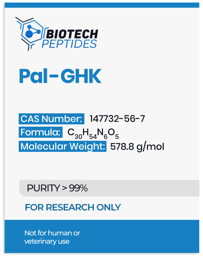
Palmitoyl Tripeptide-1, aka Pal-GHK or Palmitoyl Oligopeptide, is a hybrid peptide comprising a peptide and a fatty acid end. The peptide end is the GHK-end (Glycine, histidine, and lysine). Meanwhile, the fatty acid end is the Pal-end (Palmitoyl). Pal-GHK is a fibroblast stimulant and a small fraction of the elastin protein. It may potentially enhance the synthesis of elastin, collagen, and other extracellular matrix protein (ECM) in the connective tissues, bone, and skin.[1]

The peptide (GHK) component of the Palmitoyl Tripeptide-1 peptide may subject fibroblasts into perceiving that elastin—one of the most abundant proteins in the ECM—is damaged. As a result, the organism may rapidly produce and multiply fibroblasts to replenish and regenerate the lost elastin. Palmitoyl Tripeptide-1 may also mitigate or prevent cell aging and re-energize fibroblasts in the ECM.

The fatty acid (Palmitoyl) end of Pal-GHK appears to serve as an intermediary, a transport complex that may perform at a higher level of impact when the GHK-end is attached to the Pal-end, resulting in potentially enhanced skin and cell penetration.

Palmitoyl Tripeptide-1 may stimulate genes that might alter and reset cells. This may potentially be possible by attaching Palmitoyl to the peptide sequence, GHK, which may make it overly effective for DNA repair genes and increases expression of the 14 genes that modulate antioxidant production.[2] The researchers also note that “The Broad Institute’s Connectivity Map indicated that GHK induces a 50% or greater change of expression in 31.2% of … genes.” After the genetic changes, reduced action of cell aging and removing radicals and toxic agents that induce the development of age-related diseases might be attained.

Palmitoyl Tripeptide-1 may potentially aid in rapid tissue repair by enhancing the production of new blood vessels (angiogenesis) at injury sites. The peptide might also repair nerves and induces the growth of new nerves. Furthermore, Pal-GHK may better the actions of genes responsible for wound healing; it may activate DNA repair genes and suppresses genes that may aid cancer growth.

Scientific research has suggested that Palmitoyl Tripeptide-1 may protect the ECM by preventing cell death in certain cases. The peptide may potentiate this action by enhancing elastin and collagen production.

Palmitoyl Tripeptide-1 may potentially replenish the skin’s extracellular matrix, inducing wrinkle reduction and alleviation. At the same time, it may prevent collagen degradation following exposure to Ultraviolet A (UVA) rays.


Disclaimer: The products mentioned are not intended for human or animal consumption. Research chemicals are intended solely for laboratory experimentation and/or in-vitro testing.  Bodily introduction of any sort is strictly prohibited by law.  All purchases are limited to licensed researchers and/or qualified professionals. All information shared in this article is for educational purposes only.


References


  1. Goldsberry, S. & Garcines, L. Anti-aging cosmeceutical composition. (2013).
  2. Pickart, L., Vasquez-Soltero, J. M. & Margolina, A. GHK-Cu may Prevent Oxidative Stress in Skin by Regulating Copper and Modifying Expression of Numerous Antioxidant Genes. Cosmetics 2, 236–247 (2015). doi: 10.3390/cosmetics2030236
  3. Trookman, N. S., Rizer, R. L., Ford, R., Mehta, R. & Gotz, V. Clinical assessment of a combination lip treatment to restore moisturization and fullness. J. Clin. Aesthetic Dermatol. 2, 44–48 (2009).
  4. Dupont, E. et al. Clinical efficacy of a serum integrating multiple cosmetic ingredients in the management of erythema of the face in aging skin. J. Cosmet. Dermatol. 11, 207–212 (2012).
  5. Bradley, E. J., Griffiths, C. E. M., Sherratt, M. J., Bell, M. & Watson, R. E. B. Over-the-counter anti-ageing agents and their ability to protect and repair photoaged skin. Maturitas 80, 265–272 (2015).