Insulin-like Growth Factor 1 (IGF-1)
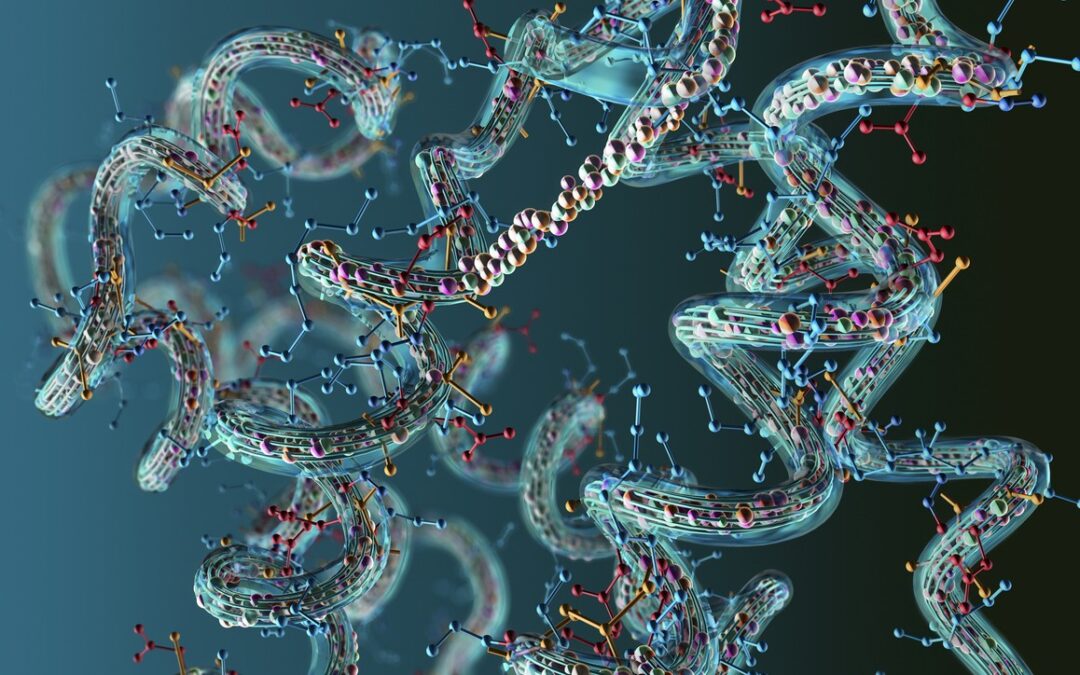
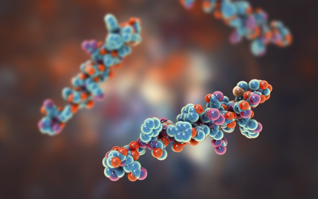
The Structure of IGF-1
The peptide consists of 70 amino acids with a molecular weight of 7649 Da. The disulfide bonds of IGF-1 may link the alpha and beta chains. Rat IGF-1 is suggested to synthesize as four precursor isoforms with alternating N-terminal and C-terminal propeptides. Mature IGF-1 is speculated to be identical between isoforms, and cleavage of the propeptide terminal is considered to produce it. There are 12 amino acids in the C-peptide region.
Mechanism of action studies on laboratory animals have suggested that Insulin-like Growth Factor 1 may be a significant mediator of growth hormone action. The pituitary gland is considered to produce growth hormone (GH), which circulates in the bloodstream, delivering signals to the liver to produce IGF-1.
Insulin-like Growth Factor 1 is speculated to bind to at least two of the receptors of cell membrane tyrosine kinases, including the IGF-1 receptor and the insulin receptor. Its main action, researchers suggest, is binding to the IGF-1 receptor, which may be ubiquitous in tissues. This ligand-receptor complex might stimulate cell growth and proliferation and emit intracellular messages via the AKT pathway known to inhibit programmed cell death. Insulin may also potentially bind to the IGF-1 receptor. Still, its affinity is suggested to be much lower, and Insulin-like Growth Factor 1 may also bind to the insulin receptor to produce a 10% effect on insulin’s potency. Researchers propose that Insulin-like Growth Factor 1 may interact with all seven IGF-1 binding proteins, specifically IGFBP2 and IGFBP5, and these two serum levels might be inversely proportional to circulating IGF-1.
Blood-transported IGF-1 has been speculated by research teams to regulate balanced growth across many tissues and organs. Stimulation of autocrine or paracrine IGF-1 might cause excessive growth as it works unaffected by growth hormones.
Insulin-like Growth Factor-1 Potential Effects
The first recorded action of the growth-stimulating effect of extrinsic IGF-1, researchers suggest, occurred by exposing a purified hormone to hypophysectomy rats.
- In diabetic rats, it may be suggested to help control glucose, and efficacy might be inversely proportional to the duration of diabetes.
- Exposure to IGF-1 may exhibit anabolic effects with subsequent rapid growth in neonatal rats, but there may be evidence that complete expression at a given concentration requires nutrition.
- Exposing animals to Insulin-like Growth Factor 1 might induce hepatoprotective and antifibrinolytic effects in experimental cirrhosis. These effects could be associated with decreased liver levels of several factors involved in oxidative damage, such as myeloperoxidase and nitric oxide.
- Obstruction of the right middle cerebral artery, researchers propose, induces ischemia, and IGF-1 expression in the central nervous system may reduce significantly. The potential exposure to Insulin-like Growth Factor 1 could increase expression in the affected muscles, sciatic nerve, lumbar spinal cord, and motor cortex. This exposure might reduce neuronal apoptosis and improve motor function.
- Immunohistochemical analysis of rat testes of various ages might reveal increased IGF-1 receptors from birth to 20 days after delivery. Tests suggest that IGF reduces Leydig cell apoptosis at all stages of development.
Disclaimer: The products mentioned are not intended for human or animal consumption. Research chemicals are intended solely for laboratory experimentation and/or in-vitro testing. Bodily introduction of any sort is strictly prohibited by law. All purchases are limited to licensed researchers and/or qualified professionals. All information shared in this article is for educational purposes only.