Sermorelin Peptide Potential as a Growth Hormone Secretagogue
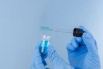
Featured Product
")
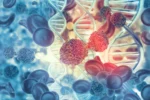
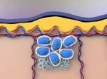
What is the Sermorelin?
Sermorelin was first synthesized in the early 1980s and has been avidly researched since its development. It is made of 29 amino acids and bears the sequence YADAXFXNSYRKVLGQLSARKLLQDXMSR. Sermorelin peptide, aka GRF (1-29), should not be confused with Modified GRF (1-29), which has 4 of the original 29 amino acids replaced with the intent to increase its half-life from 10 to 30 minutes.
General Research in Sermorelin
Sermorelin Peptide and the Endocrine System
As a GHRH analog, Sermorelin peptide has been researched primarily for its potential to stimulate pulsatile hGH synthesis via the pituitary gland. Studies observed that as long as the pituitary gland is functioning correctly, Sermorelin, combined with the amino acid Arginine, may induce a significant spike in serum HGH levels.[1] Studies in research models of growth failure also report that Sermorelin peptide appeared to increase growth velocity with growth failure and functional pituitary glands by 74%.[2] Further research reported that 6 months of either Sermorelin peptide or hGH exposure, appeared to exhibit a similar increase in growth velocity.[3] Studies also reported an apparent significant increase in serum HGH levels after several days of Sermorelin peptide introduction.[4]
The peptide may also affect the levels of other hormones apart from HGH, such as insulin and sex hormones. Although high hGH is often associated with increased insulin levels and insulin resistance, one 16-week trial suggested that Sermorelin may improve insulin sensitivity in males but not in female organisms.[5] One animal trial reported an increase in gonadotropic hormones and testosterone levels in male mice, but there is no clinical research to support these findings.[6]
Sermorelin Studies in Cardiovascular Function
Sermorelin may impact cardiovascular functioning due to its apparent HGH-stimulating potential.[7] Researchers suggest, “That GHRH analog [exposure] induced anabolic effects favoring [male species] more than [female]. Further studies are needed to define the gender differences observed in response to GHRH analog [exposure].”[8]
Sermorelin Peptide and Extracellular Matrix
Scientists posit that Sermorelin has the potential to function in increasing HGH levels. From this assumption, it may be possible for Sermorelin peptide to increase the proliferation of new skin cells, ultimately leading to increased collagen deposition inside the derma.
One study focusing on male species reported that 4 months of Sermorelin peptide exposure appeared to significantly increase their skin thickness.[11] Additionally, the males, but not the female organisms, also expressed increased libido and related behaviors.
Sermorelin Peptide and the Nervous System
One study suggested that even a one-time exposure to Sermorelin peptide may support improvements in short-term memory.[12] In fact, researchers reported that the effect on memory recall appeared more significant in Sermorelin-exposed research models than those given a placebo. There is also mixed data regarding the potential of Sermorelin peptide on brain tumors. One experiment reports that Sermorelin peptide may speed up the development of neuroendocrine tumors such as pituitary adenomas due to its possible growth-promoting effect.[13] However, a study in gliomas reported the opposite effect, observing that the peptide appeared to suppress the recurrence of the tumors.[14]
Conclusion
Sermorelin may hold significant research potential as a Growth Hormone Secretagogue (GSH). This is supported by research findings indicating that when exposed to Sermorelin, animal research models’ serum growth hormone levels might not exceed the physiological limits exerted by somatostatin.
Disclaimer: The products mentioned are not intended for human or animal consumption. Research chemicals are intended solely for laboratory experimentation and/or in-vitro testing. Bodily introduction of any sort is strictly prohibited by law. All purchases are limited to licensed researchers and/or qualified professionals. All information shared in this article is for educational purposes only.
References
- Prakash A, Goa KL. Sermorelin: a review of its use in the diagnosis and treatment of children with idiopathic growth hormone deficiency. BioDrugs. 1999 Aug;12(2):139-57. doi: 10.2165/00063030-199912020-00007. PMID: 18031173.
- Thorner M, Rochiccioli P, Colle M, Lanes R, Grunt J, Galazka A, Landy H, Eengrand P, Shah S. Once daily subcutaneous growth hormone-releasing hormone therapy accelerates growth in growth hormone-deficient children during the first year of therapy. Geref International Study Group. J Clin Endocrinol Metab. 1996 Mar;81(3):1189-96. doi: 10.1210/jcem.81.3.8772599. PMID: 8772599.
- Neyzi O, Yordam N, Ocal G, Bundak R, Darendeliler F, Açikgöz E, Berberoğlu M, Günöz H, Saka N, Calikoğlu AS. Growth response to growth hormone-releasing hormone(1-29)-NH2 compared with growth hormone. Acta Paediatr Suppl. 1993 Mar;388:16-21; discussion 22. doi: 10.1111/j.1651-2227.1993.tb12828.x. PMID: 8329826.
- Achermann JC, Hindmarsh PC, Robinson IC, Matthews DR, Brook CG. The relative roles of continuous growth hormone-releasing hormone (GHRH(1-29)NH2) and intermittent somatostatin(1-14)(SS) in growth hormone (GH) pulse generation: studies in normal and post cranial irradiated individuals. Clin Endocrinol (Oxf). 1999 Nov;51(5):575-85. doi: 10.1046/j.1365-2265.1999.00839.x. PMID: 10594518.
- Khorram O, Laughlin GA, Yen SS. Endocrine and metabolic effects of long-term administration of [Nle27]growth hormone-releasing hormone-(1-29)-NH2 in age-advanced men and women. J Clin Endocrinol Metab. 1997 May;82(5):1472-9. doi: 10.1210/jcem.82.5.3943. PMID: 9141536.
- Chatelain PG, Sanchez P, Saez JM. Growth hormone and insulin-like growth factor I treatment increase testicular luteinizing hormone receptors and steroidogenic responsiveness of growth hormone deficient dwarf mice. Endocrinology. 1991 Apr;128(4):1857-62. doi: 10.1210/endo-128-4-1857. PMID: 2004605.
- Khorram O, Laughlin GA, Yen SS. Endocrine and metabolic effects of long-term administration of [Nle27]growth hormone-releasing hormone-(1-29)-NH2 in age-advanced men and women. J Clin Endocrinol Metab. 1997 May;82(5):1472-9. doi: 10.1210/jcem.82.5.3943. PMID: 9141536.
- Vittone J, Blackman MR, Busby-Whitehead J, Tsiao C, Stewart KJ, Tobin J, Stevens T, Bellantoni MF, Rogers MA, Baumann G, Roth J, Harman SM, Spencer RG. Effects of single nightly injections of growth hormone-releasing hormone (GHRH 1-29) in healthy elderly men. Metabolism. 1997 Jan;46(1):89-96. doi: 10.1016/s0026-0495(97)90174-8. PMID: 9005976.
- Corpas E, Harman SM, Piñeyro MA, Roberson R, Blackman MR. Growth hormone (GH)-releasing hormone-(1-29) twice daily reverses the decreased GH and insulin-like growth factor-I levels in old men. J Clin Endocrinol Metab. 1992 Aug;75(2):530-5. doi: 10.1210/jcem.75.2.1379256. PMID: 1379256.
- Vittone J, Blackman MR, Busby-Whitehead J, Tsiao C, Stewart KJ, Tobin J, Stevens T, Bellantoni MF, Rogers MA, Baumann G, Roth J, Harman SM, Spencer RG. Effects of single nightly injections of growth hormone-releasing hormone (GHRH 1-29) in healthy elderly men. Metabolism. 1997 Jan;46(1):89-96. doi: 10.1016/s0026-0495(97)90174-8. PMID: 9005976.
- Khorram O, Laughlin GA, Yen SS. Endocrine and metabolic effects of long-term administration of [Nle27]growth hormone-releasing hormone-(1-29)-NH2 in age-advanced men and women. J Clin Endocrinol Metab. 1997 May;82(5):1472-9. doi: 10.1210/jcem.82.5.3943. PMID: 9141536.
- Alvarez XA, Cacabelos R. Effects of GRF (1-29) NH2 on short-term memory: neuroendocrine and neuropsychological assessment in healthy young subjects. Methods Find Exp Clin Pharmacol. 1990 Sep;12(7):493-9. PMID: 2087150.
- Stepień T, Sacewicz M, Lawnicka H, Krupiński R, Komorowski J, Siejka A, Stepień H. Stimulatory effect of growth hormone-releasing hormone (GHRH(1-29)NH2) on the proliferation, VEGF and chromogranin A secretion by human neuroendocrine tumor cell line NCI-H727 in vitro. Neuropeptides. 2009 Oct;43(5):397-400. doi: 10.1016/j.npep.2009.08.005. Epub 2009 Sep 10. PMID: 19747727.
- Chang Y, Huang R, Zhai Y, Huang L, Feng Y, Wang D, Chai R, Zhang W, Hu H. A potentially effective drug for patients with recurrent glioma: sermorelin. Ann Transl Med. 2021 Mar;9(5):406. doi: 10.21037/atm-20-6561. PMID: 33842627; PMCID: PMC8033379.
")
")
")
")