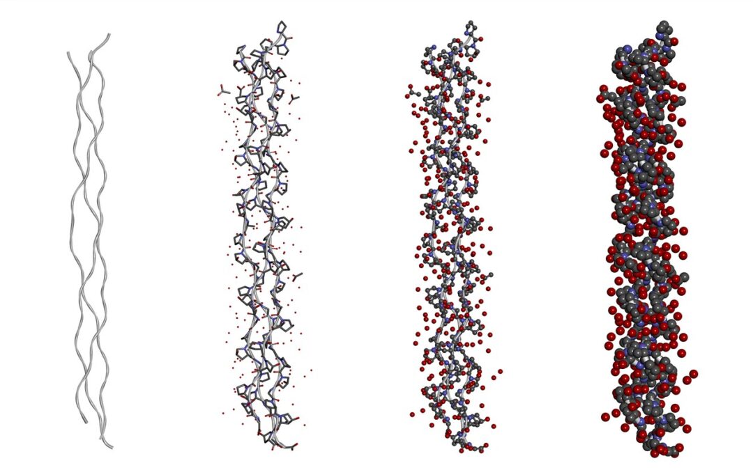
IGF-1 LR3 Peptides and Tissue Growth
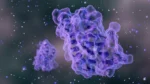
Scientists have created IGF-1 LR3 peptides by modifying the amino acid sequence of IGF-1. The new peptide has similar potential but possibly higher potency and improved stability. The LR3 in the name describes the two modifications to the IGF-1 molecule. Long R3 IGF-1, or IGF-1 LR3, is an analog of insulin-like growth factor 1 (IGF-1). IGF-1 is an anabolic peptide hormone naturally produced. Primarily, it is considered a mediator of the anabolic effects of growth hormone (HGH). Growth hormone appears to stimulate the production of IGF-1. The IGF-1 produced in the liver is released in the bloodstream and exerts anabolic effects, while the IGF-1 produced in all other tissues acts locally to stimulate growth. The first is R3 which describes the replacement of the 3rd amino acid in IGF-1 with arginine. Scientists ascribed an additional 13 amino acids to the N-terminus of the R3 IGF-1 molecule, turning it into Long R3 IGF-1. IGF-1 LR3 is an experimental peptide with potential anabolic and mitogenic effects.
IGF-1 LR3
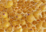
Scientists developed IGF-1 analogs such as IGF-1 LR3 primarily for experiments to stimulate cell growth. IGF-1 and its analogs appear to be highly anabolic towards actively proliferating cells. They may speed up cell replication, ultimately shortening the time required to create a cell culture and use it to conduct laboratory studies.
IGF-1 LR3 works by activating IGF-1 receptors in most animal cells. Activating these anabolic receptors increases protein synthesis and tissue growth. IGF-1 LR3 peptide appears to bind to a much lesser degree to IGF-1 proteins than other available analogs. Therefore, its affinity to the anabolic IGF-1 receptors may be much higher, and animal studies show that continuous exposure to IGF-1 LR3 leads to a 2-fold higher anabolic effect than IGF-1. Researchers also noted that “LR3 IGF-I remained more potent than IGF-I in several of these effects even when the peptides were given.“[1]
The reduced affinity to IGF-1 binding proteins may also result in a shorter half-life of IGF-1 LR3 peptide. Animal suggest show that IGF-1 LR3 is eliminated within 4 hours.[2] At the same time, it may induce higher weight gain and organ mass increase in the tested animals compared to IGF-1.
The metabolites of IGF-1 LR3 may have a longer half-life and might remain detectable in test animals for up to 16 hours. In comparison, a high percentage of the natural IGF-1 appears to bind to IGF-1 binding proteins, which may reduce its effectiveness and prolongs half-life by up to 15 hours.[3]
According to one study in a mice model of muscular dystrophy, IGF-1 LR3peptide exposure appeared highly effective in reducing contraction-mediated injury.[4] Contraction damage is a major contributing factor to the pathophysiology of muscular dystrophy.
Another animal trial by Hill et al. also suggested that an IGF-1 LR3 exposure for 8 hours may have a protective effect against loss of muscle mass loss and muscle catabolism during periods of restricted energy intake and caloric deficit. The researchers reported that “Long(R3)-IGF-1 [exposure] tended to preserve whole-body and muscle protein in beef heifers on a low-quality diet.” The continuous exposure also led to significant suppression of the natural IGF-1 synthesis.[5]
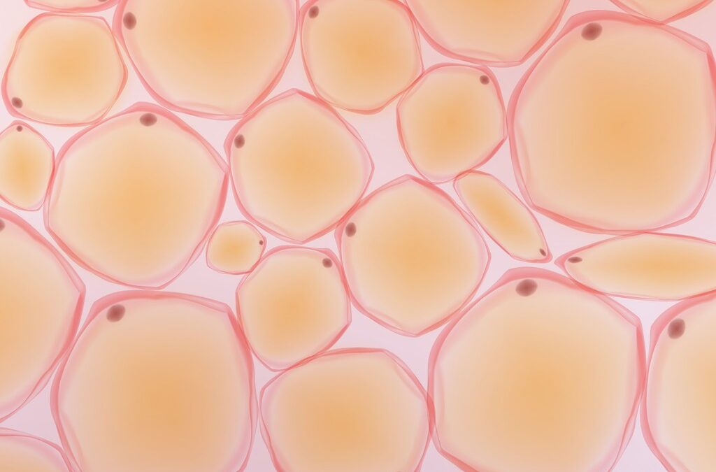
One trial in guinea pigs also reported a significant increase in the organ weight of the animals after a 7-day exposure of IGF-1 LR3. The researchers note that the exposure most significantly enlarged the adrenals, gut, kidneys, and spleen.[6] It is important to note that IGF-1 LR3 and all other IGF-1 analogs are only hypothesized to mediate the anabolic effects of growth hormone.
Disclaimer: The products mentioned are not intended for human or animal consumption. Research chemicals are intended solely for laboratory experimentation and/or in-vitro testing. Bodily introduction of any sort is strictly prohibited by law. All purchases are limited to licensed researchers and/or qualified professionals. All information shared in this article is for educational purposes only.
References
- Tomas FM, Lemmey AB, Read LC, Ballard FJ. Superior potency of infused IGF-I analogues which bind poorly to IGF-binding proteins is maintained when administered by injection. J Endocrinol. 1996 Jul;150(1):77-84. DOI: 10.1677/joe.0.1500077. PMID: 8708565.
- Mongongu C, Coudoré F, Domergue V, Ericsson M, Buisson C, Marchand A. Detection of LongR3 -IGF-I, Des(1-3)-IGF-I, and R3 -IGF-I using immunopurification and high resolution mass spectrometry for antidoping purposes. Drug Test Anal. 2021 Jul;13(7):1256-1269. DOI: 10.1002/dta.3016. Epub 2021 Feb 22. PMID: 33587816.
- Guler HP, Zapf J, Schmid C, Froesch ER. Insulin-like growth factors I and II in healthy man. Estimations of half-lives and production rates. Acta Endocrinol (Copenh). 1989 Dec;121(6):753-8. doi: 10.1530/acta.0.1210753. PMID: 2558477.
- Gehrig SM, Ryall JG, Schertzer JD, Lynch GS. Insulin-like growth factor-I analogue protects muscles of dystrophic MDX mice from contraction-mediated damage. Exp Physiol. 2008 Nov;93(11):1190-8. Doi: 10.1113/expphysiol.2008.042838. Epub 2008 Jun 20. PMID: 18567600.
- Hill RA, Hunter RA, Lindsay DB, Owens PC. Action of long(R3)-insulin-like growth factor-1 on protein metabolism in beef heifers. Domest Anim Endocrinol. 1999 May;16(4):219-29. doi: 10.1016/s0739-7240(99)00015-6. PMID: 10370861.
- Conlon MA, Tomas FM, Owens PC, Wallace JC, Howarth GS, Ballard FJ. Long R3 insulin-like growth factor-I (IGF-I) infusion stimulates organ growth but reduces plasma IGF-I, IGF-II and IGF binding protein concentrations in the guinea pig. J Endocrinol. 1995 Aug;146(2):247-53. DOI: 10.1677/joe.0.1460247. PMID: 7561636.
- Kovacs GT, Worgall S, Schwalbach P, Steichele T, Mehls O, Rosivall L. Hypoglycemic effects of insulin-like growth factor-1 in experimental uremia: can concomitant growth hormone administration prevent this effect? Horm Res. 1999;51(4):193-200. doi: 10.1159/000023357. PMID: 10474022.
- MacDonald RS. The role of insulin-like growth factors in small intestinal cell growth and development. Horm Metab Res. 1999 Feb-Mar;31(2-3):103-13. DOI: 10.1055/s-2007-978706. PMID: 10226789.
- Dunaiski V, Dunshea FR, Walton PE, Goddard C. Long [R3] insulin-like growth factor-I reduces growth, plasma growth hormone, IGF binding protein-3 and endogenous IGF-I concentrations in pigs. J Endocrinol. 1997 Dec;155(3):559-65. DOI: 10.1677/joe.0.1550559. PMID: 9488001.
- Martha S, Pantam N, Thungathurthi S, Rao VL, Devarakonda K. Study of insulin resistance in relation to serum IGF-I levels in subjects with different degrees of glucose tolerance. Int J Diabetes Dev Ctries. 2008 Apr;28(2):54-9. DOI: 10.4103/0973-3930.43100. PMID: 19902049; PMCID: PMC2772007.
")
 (200mg)")