Kisspeptin-10 Peptide: Neuroprotection, Hormonal and Reproductive Regulation

Kisspeptin-10 Peptide: Neuroprotection, Hormonal and Reproductive Regulation

Kisspeptin-10, an endogenously occurring peptide, is derived from the KISS1 gene. The original 145-amino acid polypeptide undergoes proteolytic cleavage to yield a smaller peptide comprised of 54 amino acids. This 54-amino acid peptide is further processed into Kisspeptin 45-54, commonly referred to as Kisspeptin-10.[1,2]

The KISS1 gene has been identified as a potential suppressor of metastasis in melanomas and breast carcinomas. This may suggest potential impacts on the inhibition of abnormal cell proliferation.[1] Initially recognized for its potential in metastasis suppression, subsequent research proposed that Kisspeptin-10 may hypothetically impact the hypothalamus and pituitary gland. This may imply some impact on the regulation of systems related to reproduction. Independent studies conducted in the mid-2000s suggest it may be possible for Kisspeptin-10 to be involved in hypogonadotropic hypogonadism. This may be particularly true in its capacity as a ligand for the G-protein coupled receptor 54 (GPR54).[3]


Mechanism of Action

Hypogonadotropic hypogonadism is characterized by inadequate or absent sex hormone production. This may be due to dysfunction in the pituitary gland, hypothalamus, or other parts of the brain. Gonadotropin-releasing hormone (GnRH) is considered to play a pivotal role in stimulating the pituitary gland to release follicle-stimulating hormone (FSH) and luteinizing hormone (LH), both of which are integral to various reproductive functions. Deficiencies in GnRH, FSH, and LH are identified as key contributors to hypogonadotropic hypogonadism.[4]

The GPR54 receptor also referred to as the KISS1 receptor (KISS1R), has been identified as a critical GnRH receptor.[4] Kisspeptin-10 is hypothesized to bind to GPR54 receptors, which may potentially activate the reproductive axis through the stimulation of GnRH and gonadotropin neurons.[5]

Research has isolated smaller peptide fragments, such as Kisspeptin-10, Kisspeptin-13, and Kisspeptin-14, which may exhibit biological activity towards GPR54. These peptides are proposed to bind with low affinity to GPR54 receptors, which may induce calcium mobilization, arachidonic acid release, and extracellular protein kinase phosphorylation. Such events may eventually lead to the depolarization of Kisspeptin-10 neurons and may ultimately contribute to gonadotropin release.

Ongoing research has posited several potential mechanisms of action for Kisspeptin-10. This includes the peptide’s possible role in stimulating GnRH release. The peptide may support endogenous gonadotropin release in animal models with reduced fertility. Also, the peptide may induce desensitization and suppression of the hypothalamus-pituitary-gonadal axis under certain conditions.[6]


Scientific and Research Studies


Kisspeptin-10 Peptide and Delayed Hormonal Development

The primary aim of this referenced scientific study[7] was to investigate the observed impacts of Kisspeptin-10 peptide in research models with delayed hormonal development.

In this study, researchers hypothesized that Kisspeptin-10 may stimulate gonadal hormone release and modulate reproductive function in laboratory test models. The study involved the random introduction of either Kisspeptin-10 or gonadotropin-releasing hormone (GnRH) to the test models.

Luteinizing hormone (LH) levels were monitored overnight following the introduction. Subsequently, all test models were exposed to GnRH for six days, after which LH levels were re-evaluated. The results observed by researchers indicated that 47% of the experimental group exposed to Kisspeptin-10 exhibited increased LH levels. An additional 6% of the group exhibited an intermediate response, while the remaining 47% indicated no change after exposure to the peptide.


Kisspeptin-10 Peptide and Reproductive Regulation

A comprehensive literature review conducted in 2017 examined articles published between 1999 and 2016. This review suggested that experimental data might support the hypothesis that the Kisspeptin-10 system—including the KISS1 gene and its associated GPR54 receptors—might potentially regulate the release of gonadotropin hormones. Researchers suggest that “Kisspeptin or its receptor represents a potential therapeutic target in… [test models] with fertility disorders.”[8]

Studies conducted on experimental animal models suggested that reproductive disorders like HH and PCOS might be linked to abnormalities within the KISS1 and GPR54 systems. The findings of this literature review indicated that Kisspeptin-10 may potentially function as a neuropeptide regulator of GnRH release.


Kisspeptin-10 Peptide and Reproductive Hormone Release

The objective of the study[9] was to explore the potential influence of Kisspeptin-10 on the secretion of reproductive hormones. Kisspeptin-10 was introduced to both male and female test models. The findings indicate that in male test models, exposure to the peptide may lead to an elevation in follicle-stimulating hormone (FSH) and luteinizing hormone (LH) levels. In contrast, female test models did not exhibit significant changes in FSH and LH levels throughout the menstrual cycle. Comparatively, there was a notable increase in the hormone levels of observed female test models during the preovulatory phase.


Kisspeptin-10 and Its Influence on Food Intake

Kisspeptin-10 appears to be widely distributed across various brain regions, including the hippocampus, cerebellum, posterior hypothalamus, and septum. Its presence in key nuclei involved in food intake regulation, such as the arcuate nucleus (Arc) within the hypothalamus, prompted an investigation into its potential impacts on the feeding behavior of murine test models.

An experiment[10] involving adult male murine models, aged 6 to 8 weeks and maintained under standard conditions, assessed the impact of Kisspeptin-10 on food intake. Following an overnight fast, the murine models were exposed to varying concentrations of Kisspeptin-10 or a placebo alongside a standard rodent diet and water.

Results from this study suggest that Kisspeptin-10 introduction in overnight-fasted mice led to a reduction in caloric intake during the initial 3-to-12-hour period. However, caloric intake reportedly increased during the subsequent 12-to-16-hour period, eventually aligning with levels observed in the placebo group of laboratory test models. This suggests that Kisspeptin-10 may “be a negative central regulator of feeding by increasing satiety.” Scientists observed that exposure to the peptide may lead to increased intervals between periods of calorie consumption without significant change to caloric intake.

Further research[11] has explored Kisspeptin-10’s potential impacts on caloric intake regulation within the central nervous system (CNS). Investigations have suggested that the peptide may influence the expression of genes associated with neuropeptide Y (NPY) and brain-derived neurotrophic factor (BDNF). Additionally, Kisspeptin-10 may affect neurotransmitter concentrations, including dopamine, norepinephrine, serotonin (5-hydroxytryptamine or 5-HT), dihydroxyphenylacetic acid, and 5-hydroxyindoleacetic acid in hypothalamic cells (specifically Hypo-E22 cells). Observations indicate that Kisspeptin-10 might support NPY gene expression while suppressing BDNF expression.

The peptide also appears to reduce serotonin and dopamine levels, with norepinephrine concentrations remaining stable. Notably, this reduction in dopamine and serotonin was accompanied by increased ratios of their metabolites—dihydroxyphenylacetic acid to dopamine and 5-hydroxyindoleacetic acid to serotonin—following Kisspeptin-10 exposure. The observed changes in NPY and BDNF expression, coupled with alterations in serotonin activity, may suggest a potential role for Kisspeptin-10 in influencing hunger hormone signaling.


Kisspeptin-10 and Emotional Impacts

The study aimed to investigate the impacts of Kisspeptin-10 on limbic brain activity.[12] Neuroimaging and psychometric assessments were employed to analyze the impact of Kisspeptin-10 exposure in research models. The findings suggest that Kisspeptin-10 may have influenced limbic brain function, with data suggesting heightened responsiveness to mating and bonding stimuli.


Kisspeptin-10 and Neuroprotection

The accumulation of amyloid-beta (Aβ) and alpha-synuclein (α-syn) in cholinergic neurons is associated with damage and dysfunction in critical central nervous system structures. It is hypothesized that Kisspeptin-10 may bind to extracellular Aβ, potentially mitigating its harmful impacts. Studies[13] indicate that Kisspeptin-10 may counteract the detrimental actions of Aβ, prion protein (PrP), and Islet Amyloid Polypeptide (IAPP) without interference from antagonists of the kisspeptin receptor (GPR-54) or the neuropeptide FF (NPFF) receptor. The similarity between the non-amyloid-β component (NAC) of α-syn and the C-terminus of Aβ raises the possibility that Kisspeptin-10 might also reduce α-syn-induced toxicity in cholinergic neurons.

Research[14] involving cholinergic cells suggests that while high concentrations of Kisspeptin-10 may increase toxicity, lower concentrations might decrease toxicity associated with both wild-type and E46K mutant forms of α-syn. Computational studies support these findings, and some suggest a potentially significant interaction between Kisspeptin-10 and the C-terminal residues of α-syn. Molecular dynamics simulations indicate that the complexes formed between Kisspeptin-10 and α-syn indicate substantial stability.

Further investigation[15] has focused on whether GPR54 activation is crucial for Kisspeptin-10’s ability to bind to the C-terminal regions of α-syn. One study observed choline acetyltransferase (ChAT)-positive SH-SY5Y neurons, genetically modified to express either wild-type or E46K mutant α-syn, to evaluate the impact of Kisspeptin-10 on neuronal damage through flow cytometry and immunocytochemistry.

Some findings suggest that Kisspeptin-10 may reduce both apoptosis and mitochondrial damage in neurons affected by α-syn, with its protective impacts remaining unaffected by the presence of a GPR54 antagonist, kisspeptin-234 (KP-234). This implies that GPR54 activation might not be necessary for Kisspeptin-10’s neuroprotective impacts. Additionally, Kisspeptin-10 appears to lower the levels of α-syn and ChAT in neurons overexpressing both wild-type and E46K mutant α-syn.

Disclaimer: The products mentioned are not intended for human or animal consumption. Research chemicals are intended solely for laboratory experimentation and/or in-vitro testing. Bodily introduction of any sort is strictly prohibited by law. All purchases are limited to licensed researchers and/or qualified professionals. All information shared in this article is for educational purposes only.


References:

  1. KISS1 KiSS-1 metastasis suppressor [Homo sapiens (humans)]. https://www.ncbi.nlm.nih.gov/gene/3814
  2. Mead EJ, Maguire JJ, Kuc RE, Davenport AP. Kisspeptins: a multifunctional peptide system with a role in reproduction, cancer, and the cardiovascular system. Br J Pharmacol. 2007 Aug;151(8):1143-53. doi: 10.1038/sj.bjp.0707295. Epub 2007 May 21. PMID: 17519946; PMCID: PMC2189831. https://www.ncbi.nlm.nih.gov/pmc/articles/PMC2189831/
  3. Messager S, Chatzidaki EE, Ma D, Hendrick AG, Zahn D, Dixon J, Thresher RR, Malinge I, Lomet D, Carlton MB, Colledge WH, Caraty A, Aparicio SA. Kisspeptin directly stimulates gonadotropin-releasing hormone release via G protein-coupled receptor 54. Proc Natl Acad Sci U S A. 2005 Feb 1;102(5):1761-6. doi: 10.1073/pnas.0409330102. Epub 2005 Jan 21. PMID: 15665093; PMCID: PMC545088. https://www.ncbi.nlm.nih.gov/pmc/articles/PMC545088/
  4. Hypogonadotropic hypogonadism. US National Library of Medicine. https://medlineplus.gov/ency/article/000390.htm
  5. Rønnekleiv OK, Kelly MJ. Kisspeptin excitation of GnRH neurons. Adv Exp Med Biol. 2013;784:113-31. doi: 10.1007/978-1-4614-6199-9_6. PMID: 23550004; PMCID: PMC4019505. target=”_blank” rel=”noopener”https://www.ncbi.nlm.nih.gov/pmc/articles/PMC4019505/
  6. Prague JK, Dhillo WS. Potential Clinical Use of Kisspeptin. Neuroendocrinology. 2015;102(3):238-45. doi: 10.1159/000439133. Epub 2015 Aug 7. PMID: 26277870. https://pubmed.ncbi.nlm.nih.gov/26277870/
  7. Kristen P. Tolson et al., Impaired kisspeptin signaling decreases metabolism and promotes glucose intolerance and obesity. The Journal of Clinical Investigation. Published June 17, 2014. https://www.jci.org/articles/view/71075
  8. Zeydabadi Nejad S, Ramezani Tehrani F, Zadeh-Vakili A. The Role of Kisspeptin in Female Reproduction. Int J Endocrinol Metab. 2017 Apr 22;15(3):e44337. doi: 10.5812/ijem.44337. PMID: 29201072; PMCID: PMC5702467. https://www.ncbi.nlm.nih.gov/pmc/articles/PMC5702467/
  9. Comninos AN, Wall MB, Demetriou L, Shah AJ, Clarke SA, Narayanaswamy S, Nesbitt A, Izzi-Engbeaya C, Prague JK, Abbara A, Ratnasabapathy R, Salem V, Nijher GM, Jayasena CN, Tanner M, Bassett P, Mehta A, Rabiner EA, Hönigsperger C, Silva MR, Brandtzaeg OK, Lundanes E, Wilson SR, Brown RC, Thomas SA, Bloom SR, Dhillo WS. Kisspeptin modulates sexual and emotional brain processing in humans. J Clin Invest. 2017 Feb 1;127(2):709-719. doi: 10.1172/JCI89519. Epub 2017 Jan 23. PMID: 28112678; PMCID: PMC5272173. https://www.ncbi.nlm.nih.gov/pmc/articles/PMC5272173/
  10. Stengel, A., Wang, L., Goebel-Stengel, M., & Taché, Y. (2011). Centrally injected kisspeptin reduces food intake by increasing meal intervals in mice. Neuroreport, 22(5), 253–257. https://www.ncbi.nlm.nih.gov/pmc/articles/PMC3063509/
  11. Orlando G, Leone S, Ferrante C, Chiavaroli A, Mollica A, Stefanucci A, Macedonio G, Dimmito MP, Leporini L, Menghini L, Brunetti L, Recinella L. s of Kisspeptin-10 on Hypothalamic Neuropeptides and Neurotransmitters Involved in Appetite Control. Molecules. 2018 Nov 24;23(12):3071. Doi: 10.3390/molecules23123071. PMID: 30477219; PMCID: PMC6321454. https://www.ncbi.nlm.nih.gov/pmc/articles/PMC6321454/
  12. Chan YM, Lippincott MF, Kusa TO, Seminara SB. Divergent responses to kisspeptin in children with delayed puberty. JCI Insight. 2018 Apr 19;3(8):e99109. doi: 10.1172/jci.insight.99109. PMID: 29669934; PMCID: PMC5931121. https://www.ncbi.nlm.nih.gov/pmc/articles/PMC5931121/
  13. Milton NG, Chilumuri A, Rocha-Ferreira E, Nercessian AN, Ashioti M. Kisspeptin prevention of amyloid-β peptide neurotoxicity in vitro. ACS Chem Neurosci. 2012 Sep 19;3(9):706-19. doi: 10.1021/cn300045d. Epub 2012 May 30. PMID: 23019497; PMCID: PMC3447396. https://pubmed.ncbi.nlm.nih.gov/23019497/
  14. Simon, C., Soga, T., Ahemad, N., Bhuvanendran, S., & Parhar, I. (2022). Kisspeptin-10 Rescues Cholinergic Differentiated SHSY-5Y Cells from α-Synuclein-Induced Toxicity In Vitro. International journal of molecular sciences, 23(9), 5193. https://doi.org/10.3390/ijms23095193
  15. Simon, C., Soga, T., & Parhar, I. (2023). Kisspeptin-10 Mitigates α-Synuclein-Mediated Mitochondrial Apoptosis in SH-SY5Y-Derived Neurons via a Kisspeptin Receptor-Independent Manner. International journal of molecular sciences, 24(7), 6056. https://doi.org/10.3390/ijms24076056
  16. Image Source: https://pubchem.ncbi.nlm.nih.gov/compound/Kisspeptin-10
Vialox Peptide: Neuromuscular Transmission and Skin Wrinkle Reduction

Vialox Peptide: Neuromuscular Transmission and Skin Wrinkle Reduction

Vialox, also known as Pentapeptide-3v, is a synthetic peptide characterized by the amino acid sequence GPRPA.[1] This molecule is posited to function through the inhibition of neuronal nicotinic acetylcholine receptors located within the postsynaptic membrane of muscle cells. These receptors are deemed responsible for the transmission of signals from nerve cells to muscle cells, which subsequently results in muscle contraction.

By potentially diminishing the release of acetylcholine, Vialox may contribute to muscle relaxation, thereby possibly reducing the depth and formation of wrinkles along the skin barrier. The proposed mechanism of action for Vialox bears resemblance to that of tubocurarine, a natural alkaloid compound known for its muscle-relaxing properties.


Scientific and Research Studies


Action Mechanisms

Vialox is suggested by researchers to inhibit neurotransmitter activity.[2] It may function similarly to tubocurarine through its interaction with acetylcholine receptors on the postsynaptic membrane of muscle cells.[3] Tubocurarine, a naturally occurring alkaloid found in the bark of plants such as Chondrodendron tomentosum (commonly known as “curare”), is considered a potent neurotoxin. It acts as a non-depolarizing neuromuscular blocker by preventing acetylcholine from inducing muscle contraction at the neuromuscular junction.

Researchers also classify Vialox as a non-depolarizing neuromuscular blocker. The peptide binds to acetylcholine receptors on the postsynaptic membrane of muscle cells, acting as “a competitive antagonist” at these receptor sites.[4] It may interact with neuronal nicotinic acetylcholine receptors, which play a role in regulating muscle contraction by mediating communication between motor nerves and muscles at the neuromuscular junction.

As an antagonist, Vialox appears to block acetylcholine binding to these receptors, preventing the opening of sodium ion channels required for depolarizing the cell and initiating muscle contraction.[5] By inhibiting acetylcholine receptor activity, Vialox may potentially keep smooth muscles relaxed, possibly reducing wrinkles in the skin barrier as a result.


Vialox Peptide and Wrinkle Formation, Skin Texture

Research into Vialox has explored its potential to mitigate wrinkles on the skin surface and decrease texture variations along the skin barrier. The study of compounds in wrinkle reduction carries certain risks, particularly with higher concentrations or prolonged exposure. Long-term exposure may also pose unknown or unanticipated ancillary impacts within the laboratory test models. However, Vialox is noted for its short half-life and potential for less abrasive impacts. Studies suggest that this compound may cause “softened wrinkles and reduced skin roughness.”[5]

Experimental data indicated a reduction in muscle contractions by 71% within one minute of Vialox exposure, with a subsequent 58% reduction observed after two hours. These findings imply that the decreased frequency of muscle contractions may contribute to the formation of shallower lines on the skin’s surface.

Additional research supports Vialox’s potential in reducing the development and depth of skin wrinkles.[7] Results indicate a 49% decrease in wrinkle size and a 47% reduction in skin roughness after 28 days of consistent exposure.


Vialox Peptide and Neuromuscular Transmission

Vialox has garnered scientific interest due to its proposed ability to disrupt nerve-muscle communication. Unlike other antagonists of nicotinic acetylcholine receptors (AChR), Vialox appears to act exclusively on peripheral AChR, with minimal impact on central neuronal receptors as suggested by animal studies. Researchers hypothesize that Vialox might be effective in addressing certain spastic conditions, including migraines and muscle spasms.

Vialox is suggested to interfere with signal transmission between neurons and muscles by acting as an antagonist of the acetylcholine receptor. It appears to block nerve signals at the post-synaptic membrane, leading to muscle relaxation. Normally, when a nerve’s axon releases acetylcholine, these signals travel to the neuromuscular junction and bind to receptors on the muscle, facilitating the release of sodium ions. This process may result in depolarization, generating the electrical pulse responsible for muscle contraction and wrinkle formation.

Vialox is studied for its potential to halt this process by binding to AChR, thereby preventing acetylcholine from attaching to these receptors. This blockade is thought to reduce both the frequency and intensity of muscular contractions, similar to the effects induced by botulinum toxin, tubocurarine, and curare toxin. The consequent partial paralysis of muscles leads to forced relaxation.

Disclaimer: The products mentioned are not intended for human or animal consumption. Research chemicals are intended solely for laboratory experimentation and/or in-vitro testing. Bodily introduction of any sort is strictly prohibited by law. All purchases are limited to licensed researchers and/or qualified professionals. All information shared in this article is for educational purposes only.


References:

  1. National Center for Biotechnology Information (2024). PubChem Compound Summary for CID 67073230, Vialox. Retrieved August 4, 2024 from https://pubchem.ncbi.nlm.nih.gov/compound/Vialox.
  2. Husein el Hadmed, H., & Castillo, R. F. (2016). Cosmeceuticals: peptides, proteins, and growth factors. Journal of cosmetic dermatology, 15(4), 514-519. Husein el Hadmed, H., & Castillo, R. F. (2016). Cosmeceuticals: peptides, proteins, and growth factors. Journal of cosmetic dermatology, 15(4), 514-519. https://pubmed.ncbi.nlm.nih.gov/27142709/
  3. Lupo, M. P., & Cole, A. L. (2007). Cosmeceutical peptides. Dermatologic therapy, 20(5), 343-349. https://pubmed.ncbi.nlm.nih.gov/18045359/
  4. Gorouhi, F., & Maibach, H. I. (2009). Role of peptides in preventing or treating aged skin. International journal of cosmetic science, 31(5), 327-345. https://pubmed.ncbi.nlm.nih.gov/19570099/
  5. Satriyasa B. K. (2019). Botulinum toxin (Botox) A for reducing the appearance of facial wrinkles: a literature review of clinical use and pharmacological aspect. Clinical, cosmetic and investigational dermatology, 12, 223–228. https://doi.org/10.2147/CCID.S202919
  6. Kalandakanond, S., & Coffield, J. A. (2001). Cleavage of SNAP-25 by botulinum toxin type A requires receptor-mediated endocytosis, pH-dependent translocation, and zinc. The Journal of pharmacology and experimental therapeutics, 296(3), 980–986. https://pubmed.ncbi.nlm.nih.gov/11181932/
  7. Reddy BY, Jow T, Hantash BM. Bioactive oligopeptides in dermatology: Part II. Exp Dermatol. 2012 Aug;21(8):569-75. doi: 10.1111/j.1600-0625.2012.01527.x. Epub 2012 Jun 4. PMID: 22672721. https://pubmed.ncbi.nlm.nih.gov/22672721/
  8. Lebedev DS, Kryukova EV, Ivanov IA, Egorova NS, Timofeev ND, Spirova EN, Tufanova EY, Siniavin AE, Kudryavtsev DS, Kasheverov IE, Zouridakis M, Katsarava R, Zavradashvili N, Iagorshvili I, Tzartos SJ, Tsetlin VI. Oligoarginine Peptides, a New Family of Nicotinic Acetylcholine Receptor Inhibitors. Mol Pharmacol. 2019 Nov;96(5):664-673. doi: 10.1124/mol.119.117713. Epub 2019 Sep 6. PMID: 31492697. https://pubmed.ncbi.nlm.nih.gov/31492697/
Retatrutide Peptide, and Advances in Energy Intake and Metabolism Research

Retatrutide Peptide, and Advances in Energy Intake and Metabolism Research

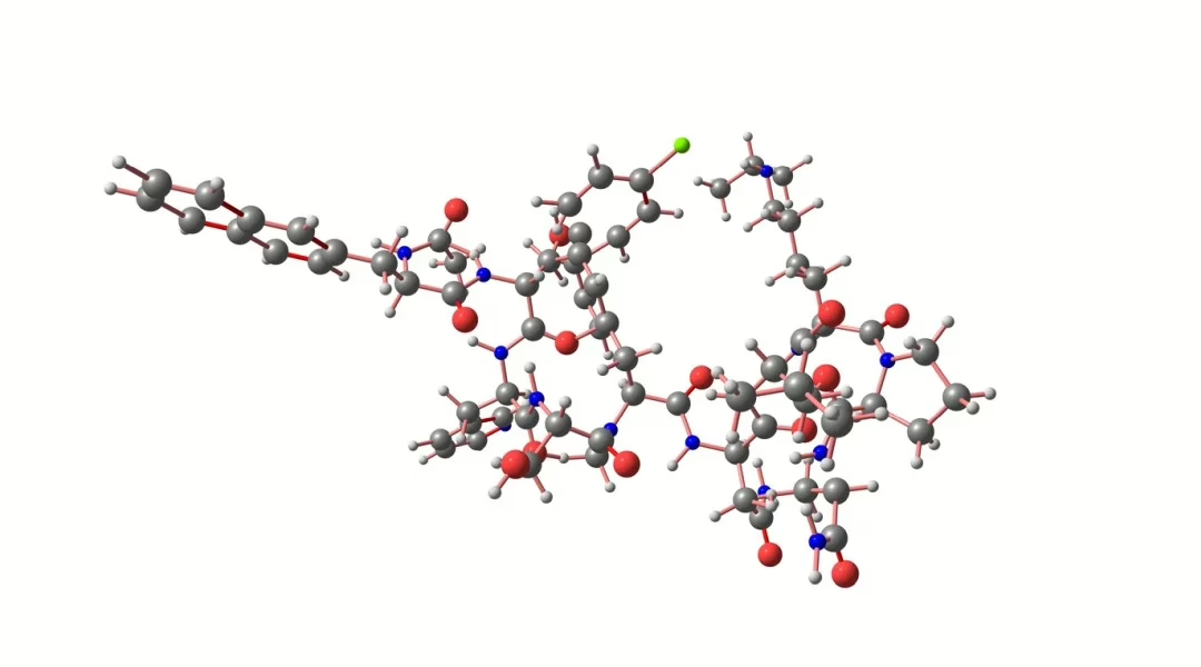
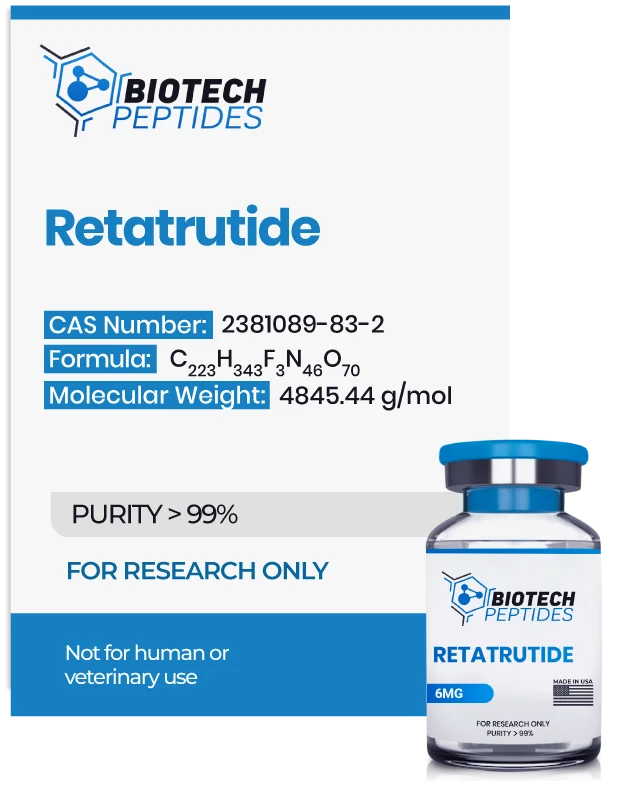
Retatrutide, also known by the identifier LY3437943, is a synthetic peptide characterized as a triple receptor agonist, comprising a sequence of 39 amino acids.[1]

Designed as an analog to gastric inhibitory polypeptide (GIP), this compound reportedly exhibits additional affinity towards the glucagon-like peptide-1 (GLP-1) receptor and the glucagon (GCG) receptor.[2] These receptors typically bind to their respective endogenous hormones, GIP, GLP-1, and GCG, which function as key hormonal regulators within the endocrine system. It is proposed that GLP-1 and GIP, acting as incretin hormones, might enhance insulin secretion from pancreatic beta cells and potentially increase satiety following nutrient intake. Conversely, glucagon is thought to play a compensatory role by potentially elevating blood glucose levels during fasting states.

Moreover, activation of GLP-1 receptors is suggested to slow gastric emptying, while stimulation of GCG receptors is hypothesized to increase energy expenditure and fat metabolism, notably influencing hepatic processes and converting white adipose tissue to beige adipose tissue, which is believed to possess thermogenic properties similar to brown adipose tissue, thereby potentially enhancing thermogenesis and metabolic rates.

The interaction of Retatrutide with these receptors indicates a multifaceted impact on metabolic regulation, which may be significant in the study of glycemic control and weight management. Additionally, Retatrutide has undergone chemical modification with a C20 moiety, purportedly extending its half-life to approximately six days.[3]


Scientific and Research Studies


Retatrutide Peptide and Glycated Hemoglobin Levels

Retatrutide has indicated potential in reducing glycated hemoglobin (HbA1c) levels in models of hyperglycemia and baseline HbA1c exceeding 7%. Data from phase 2 trials lasting up to 36 weeks suggest that Retatrutide may decrease HbA1c levels by as much as 2.16% (23.59 mmol/mol), indicating a significant amelioration of hyperglycemia. The findings also highlighted a notable reduction in total weight, with decreases from baseline to 36 weeks reaching up to a least-squares mean of 16.94% in the Retatrutide-exposed groups.[4]


Retatrutide Peptide and Appetite and Energy Intake

In a 48-week phase 2 study, Retatrutide has been investigated for its potential to significantly reduce total energy intake and facilitate the maintenance of a caloric deficit.

The research suggests that Retatrutide may lead to a substantial reduction in baseline weight, with observed decreases exceeding 24.2%, compared to a 2.1% reduction observed in the control group. Additionally, the study reported improvements in several cardiometabolic parameters, including indicators related to cholesterol metabolism, glucose regulation, and insulin sensitivity.[5]

Researchers noted that in type 2 diabetes models, Retatrutide peptide showed “meaningful improvements in glycaemic control and robust reductions in bodyweight, with a […] profile consistent with GLP-1 receptor agonists and GIP and GLP-1 receptor agonists.”


Retatrutide Peptide and GIP Receptors Interaction

Retatrutide’s mechanism of action includes the activation of gastric inhibitory polypeptide (GIP) receptors. A comprehensive study has investigated the possible intricate roles of GIP receptor activation in the regulation of energy balance. Researchers hypothesize that Retatrutide may exert influence through central neural pathways, particularly in key brain regions such as the hypothalamus and brainstem[6], which are considered critical for maintaining energy homeostasis and appetite. It is proposed that GIP receptor agonists may directly interact with neurons in these regions, potentially altering neuronal activity to reduce caloric intake and promote a negative energy balance.

The hypothalamus, a regulator in this process, contains specific nuclei—such as the arcuate, paraventricular, and ventromedial nuclei—that are believed to be essential in the regulation of appetite and energy expenditure. Activation of GIP receptors within these nuclei may suppress appetite or enhance signals of satiety. For example, GIP receptor activation in the arcuate nucleus might modulate neurons producing neuropeptide Y (NPY), which is associated with increased appetite, and pro-opiomelanocortin (POMC), which is linked to appetite suppression. This suggests that GIP receptor agonists might influence these neuropeptide systems, potentially leading to reduced caloric intake.

Moreover, the study explores the potential impact of GIP receptor agonists on the brain’s emetic centers. By potentially modulating neural circuits in the area postrema and the nucleus tractus solitarius of the brainstem—key areas involved in nausea and vomiting—GIP receptor agonists might inhibit these responses. Additionally, the role of GIP receptor agonists in enhancing the permeability of the blood-brain barrier (BBB) is proposed as a mechanism to improve the delivery and efficacy of agents targeting brain regions involved in energy balance. This may involve the modulation of tight junctions or transport systems within the neurovascular unit, facilitating increased access of these agents to the central nervous system (CNS).


Retatrutide Peptide and Energy Metabolism

Activation of glucagon (GCG) receptors by agonists such as Retatrutide may promote increased energy expenditure and fat oxidation, potentially enhancing energy utilization. Studies suggest that Retatrutide may activate GCG receptors in hepatocytes, possibly boosting energy expenditure by increasing lipid catabolism and enhancing the liver’s metabolic rate. This might be facilitated through mechanisms such as hepatic futile cycling, enhanced mitochondrial function, and the secretion of thermogenic agents such as fibroblast growth factor 21 (FGF21) and bile acids, which may further augment energy expenditure. The activation of GCG receptors in the liver by Retatrutide may trigger a cascade of metabolic events that induce weight loss, including the reduction of hepatic steatosis through increased lipid oxidation, elevated activity of metabolic enzymes, and upregulation of mitochondrial biogenesis.[7] Key hormones like FGF21 and bile acids, released by the liver in response to GCG receptor activation, might play a role in systemic energy homeostasis.

Retatrutide may also stimulate GCG receptors in adipocytes, initiating a process known as beiging, which transforms white adipocytes into beige adipocytes. Beige adipocytes, similar to brown adipocytes, may contribute to increased caloric expenditure by generating heat, potentially enhancing thermogenesis and metabolic rate. The beiging of white adipose tissue may involve the induction of UCP1-dependent non-shivering thermogenesis and metabolic futile cycles, such as the creatine and succinate cycles, which may dissipate energy as heat. Additionally, Retatrutide might upregulate the expression of thermogenic genes in both brown and beige adipocytes, promoting the formation and activity of these thermogenically active cells.

Furthermore, Retatrutide’s activation of GCG receptors may extend to enhancing the thermogenic capacity of brown adipose tissue (BAT). Activation of BAT by Retatrutide may increase metabolic rate through uncoupling protein 1 (UCP1), which may dissipate the mitochondrial proton gradient as heat. This process might utilize stored lipids and support the oxidation of circulating glucose, lipids, and amino acids, contributing to overall energy expenditure. Based on this research, the researchers propose that “the thermogenic activity of GCGR agonism” may assist “in lowering weight.”


Retatrutide Peptide and Metabolic Influence

Research posits that Retatrutide may exert its effects through the activation of glucagon-like peptide-1 (GLP-1) receptors, which are typically ubiquitous. This peptide potentially interacts with GLP-1 receptors in the pancreas, which may stimulate insulin secretion from pancreatic beta cells and reduce glucagon production from alpha cells in a glucose-dependent manner. The GLP-1 receptor, a member of the class B G protein-coupled receptor family, is thought to primarily engage the cAMP-PKA signaling pathway in the pancreas. The interaction between GLP-1 and its receptor is believed to activate adenylate cyclase (AC), which converts ATP to cyclic adenosine monophosphate (cAMP), potentially increasing cAMP levels. This elevation in cAMP might activate protein kinase A (PKA) and the guanine nucleotide exchange factor RAPGEF4 (also known as EPAC2). The activated PKA may close ATP-sensitive K+ channels and depolarize the cell membrane, possibly activating voltage-dependent Ca2+ channels, leading to Ca2+ influx and the generation of action potentials. Furthermore, PKA might facilitate the release of Ca2+ by activating inositol trisphosphate (IP3). The activated EPAC2 may also activate Ras-related protein 1 and phospholipase C, possibly engaging the IP3 and diacylglycerol (DAG) pathways to further enhance Ca2+ release. Collectively, these pathways may increase intracellular Ca2+ levels, potentially boosting mitochondrial ATP synthesis and promoting insulin secretion via exocytosis.[7]

Similar to its effects on GIP receptors, Retatrutide is suggested to influence neurons in the hypothalamic arcuate nucleus, involved in the regulation of appetite and hunger, possibly through GLP-1 receptors. These neurons express neuropeptides such as pro-opiomelanocortin (POMC) and cocaine- and amphetamine-regulated transcript (CART). It is theorized that direct activation of GLP-1 receptors on POMC/CART neurons might induce satiety and indirectly inhibit the release of hunger-stimulating peptides neuropeptide Y (NPY) and agouti-related peptide (AgRP). Research also indicates that activation of GLP-1 receptors by agonists like Retatrutide may maintain elevated levels of free leptin and peptide YY3-36 (PYY3-36) during the reduction of weight.[8]

Disclaimer: The products mentioned are not intended for human or animal consumption. Research chemicals are intended solely for laboratory experimentation and/or in-vitro testing. Bodily introduction of any sort is strictly prohibited by law. All purchases are limited to licensed researchers and/or qualified professionals. All information shared in this article is for educational purposes only.


References:

  1. Folli F, Finzi G, Manfrini R, Galli A, Casiraghi F, Centofanti L, Berra C, Fiorina P, Davalli A, La Rosa S, Perego C, Higgins PB. Mechanisms of action of incretin receptor based dual- and tri-agonists in pancreatic islets. Am J Physiol Endocrinol Metab. 2023 Nov 1;325(5):E595-E609. doi: 10.1152/ajpendo.00236.2023. Epub 2023 Sep 20. PMID: 37729025; PMCID: PMC10874655. https://www.ncbi.nlm.nih.gov/pmc/articles/PMC10874655/
  2. Jakubowska A, Roux CWL, Viljoen A. The Road towards Triple Agonists: Glucagon-Like Peptide 1, Glucose-Dependent Insulinotropic Polypeptide and Glucagon Receptor – An Update. Endocrinol Metab (Seoul). 2024 Feb;39(1):12-22. doi: 10.3803/EnM.2024.1942. Epub 2024 Feb 14. PMID: 38356208; PMCID: PMC10901658. https://pubmed.ncbi.nlm.nih.gov/38356208/
  3. Doggrell SA. Is retatrutide (LY3437943), a GLP-1, GIP, and glucagon receptor agonist a step forward in the treatment of diabetes and obesity? Expert Opin Investig Drugs. 2023 May;32(5):355-359. doi: 10.1080/13543784.2023.2206560. Epub 2023 Apr 24. PMID: 37086147. https://pubmed.ncbi.nlm.nih.gov/37086147/
  4. Rosenstock J, Frias J, Jastreboff AM, Du Y, Lou J, Gurbuz S, Thomas MK, Hartman ML, Haupt A, Milicevic Z, Coskun T. Retatrutide, a GIP, GLP-1 and glucagon receptor agonist, for people with type 2 diabetes: a randomised, double-blind, placebo and active-controlled, parallel-group, phase 2 trial conducted in the USA. Lancet. 2023 Aug 12;402(10401):529-544. doi: 10.1016/S0140-6736(23)01053-X Epub 2023 Jun 26. PMID: 37385280. https://www.thelancet.com/journals/lancet/article/PIIS0140-6736(23)01053-X/abstract
  5. Jastreboff AM, Kaplan LM, Frías JP, Wu Q, Du Y, Gurbuz S, Coskun T, Haupt A, Milicevic Z, Hartman ML; Retatrutide Phase 2 Obesity Trial Investigators. Triple-Hormone-Receptor Agonist Retatrutide for Obesity – A Phase 2 Trial. N Engl J Med. 2023 Aug 10;389(6):514-526. doi: 10.1056/NEJMoa2301972. Epub 2023 Jun 26. PMID: 37366315. https://pubmed.ncbi.nlm.nih.gov/37366315/
  6. Samms RJ, Sloop KW, Gribble FM, Reimann F, Adriaenssens AE. GIPR Function in the Central Nervous System: Implications and Novel Perspectives for GIP-Based Therapies in Treating Metabolic Disorders. Diabetes. 2021 Sep;70(9):1938-1944. doi: 10.2337/dbi21-0002. Epub 2021 Jun 27. PMID: 34176786; PMCID: PMC8576420. https://www.ncbi.nlm.nih.gov/pmc/articles/PMC8576420/
  7. Zhao X, Wang M, Wen Z, Lu Z, Cui L, Fu C, Xue H, Liu Y, Zhang Y. GLP-1 Receptor Agonists: Beyond Their Pancreatic Effects. Front Endocrinol (Lausanne). 2021 Aug 23;12:721135. doi: 10.3389/fendo.2021.721135. PMID: 34497589; PMCID: PMC8419463. https://www.ncbi.nlm.nih.gov/pmc/articles/PMC8419463/
  8. Ard J, Fitch A, Fruh S, Herman L. Weight Loss and Maintenance Related to the Mechanism of Action of Glucagon-Like Peptide 1 Receptor Agonists. Adv Ther. 2021 Jun;38(6):2821-2839. doi: 10.1007/s12325-021-01710-0. Epub 2021 May 11. PMID: 33977495; PMCID: PMC8189979. https://pubmed.ncbi.nlm.nih.gov/33977495/
Research in LL-37 Peptide and Immunomodulation, Disease Pathophysiology

Research in LL-37 Peptide and Immunomodulation, Disease Pathophysiology

LL-37 peptide, also recognized as Cathelicidin, appears to be a significant player in the realm of antimicrobial peptides (AMPs), characterized by its cationic nature and composed of 37 amino acids. LL-37 is primarily synthesized in neutrophils, though its presence extends to macrophages and polymorphonuclear leukocytes.[1] This cationic peptide, a member of the antimicrobial peptide (AMP) family, has garnered significant attention within scientific circles due to its potentially diverse functions.

Studies suggest that the peptide’s genesis lies in the breakdown of hCAP18 proteins by protease enzymes, giving rise to LL-37. Researchers evaluating its antimicrobial potential have that the peptide may host the ability to form agglomerates and lipid bilayers, conferring resistance against enzymatic degradation.[2] Structurally, LL-37 adopts an α-helical configuration, which is believed to be critical for its interactions with microbial membranes.

Studies[2] exploring LL-37’s mechanism of action indicate its potential in combating microbial threats. Through electrostatic interactions, LL-37 appears to interface with bacterial membranes, leading to membrane interference and eventual degradation of the bacterial cell. The peptide’s mode of action encompasses pore formation and profound disruption of lipid complexes, underscoring its potential as a defense against microbial invasion.


LL-37 Peptide and Inflammation

Research suggests that LL-37 peptide may exhibit multifaceted impact. These findings have increased speculation on the peptide’s potential to exert nuanced immune modulation. Among its suggested impacts, LL-37 appears to potentially attenuate keratinocyte apoptosis, boost IFN-alpha production, modulate chemotaxis of neutrophils and eosinophils, dampen toll-like receptor 4 (TLR4) signaling, enhance IL-18 production, and mitigate atherosclerotic plaque formation.

Speculated to have dynamic influence on the immune system, LL-37 appears to be subject to modulation by the inflammatory microenvironment. In cell culture studies, immune cell responses to LL-37 vary based on their activation status. T-cells, for instance, are suggested to exhibit heightened inflammatory responses in the presence of LL-37 when in a quiescent state,[3] yet temper their inflammatory actions upon prior activation. This nuanced interplay suggests LL-37’s homeostatic role in immune regulation, delicately balancing immune responses to prevent hyperactivation in the face of infection.

Studies discussing the role of LL-37 in the modulation of immune and inflammatory pathways suggest that elevated LL-37 levels might serve as a safeguard against exacerbated inflammation in autoimmune conditions, including “in the pathogenesis of systemic lupus erythematosus, rheumatoid arthritis, atherosclerosis, and possibly other diseases.”[4]


LL-37 Peptide and Antimicrobial Action

LL-37 is speculated to serve as a frontline defender against invading pathogens by reportedly swiftly mobilizing in response to infection. Research elucidates that while normal skin cells maintain minimal levels of LL-37, its expression appears to surge rapidly upon encountering microbial intrusion, highlighting its possible role in combating infections at the skin barrier. Furthermore, synergistic interactions with proteins such as beta-defensin 2 suggest the complexity of LL-37’s immune functions.[5]

Functionally, LL-37 appears to operate by binding to bacterial lipopolysaccharide (LPS), a crucial constituent of the outer membrane of gram-negative bacteria. The peptide’s affinity for LPS appears to disrupt the membrane’s integrity, possibly rendering it lethal against these pathogens. Consequently, there is burgeoning interest in exploring further potential of exogenous LL-37 to combat severe bacterial infections in preclinical research.[6]

Interestingly, while LL-37 is suggested to predominantly target the cell membrane components of gram-negative bacteria, its efficacy appears to extend to gram-positive counterparts as well. This broad-spectrum activity positions it as a promising candidate for addressing infections caused by staphylococcal strains and other formidable pathogens. In vitro investigations speculate that LL-37 augments the antimicrobial effects of lysozyme, accentuating its capacity to combat gram-positive bacteria like Staphylococcus aureus.[7]


LL-37 Peptide and the Intestine


Intestinal Physiology:

In investigations conducted within cell cultures, LL-37 is suggested to possess a spectrum of actions within the intestinal milieu. Primarily, the peptide is speculated to facilitate the migration of cells pivotal for the maintenance of the intestinal epithelial barrier. Additionally, LL-37 appears to exhibit a mitigating action on apoptosis amidst intestinal inflammation, thereby possibly attenuating the pathogenesis of various inflammatory conditions.[8]

In the intricate landscape of the intestine, it appears that LL-37 does not act in isolation but rather synergizes with beta-defensin 2, possibly fostering wound healing processes. Cellular studies suggest the collaborative action of these peptides in the repair and preservation of intestinal epithelium, concomitantly abating TNF-related cell death. Presently, TNF-alpha inhibitors combat inflammatory bowel diseases, albeit accompanied by notable adverse impacts, including possible elevated risk of severe infection. The emergence of LL-37-based interventions for inflammatory bowel diseases may mitigate reliance on TNF-alpha inhibitors.


Intestinal Malignancies:

Exploration into the interplay between LL-37 and cancer yields heterogeneous findings, yet evidences its potential action in intestinal and gastric malignancies, including squamous cell carcinoma. Interestingly, these hypothesized actions appear to be modulated via a vitamin D-dependent pathway, suggesting that this “activates the anti-cancer activity of tumor-associated macrophages (TAMs) and enhances antibody-dependent cellular cytotoxicity (ADCC)” speculated to support the “critical roles of vitamin D-dependent induction of cathelicidin in cancer progression.”[9]


LL-37 Peptide and Arthritic Pathophysiology

Research conducted in rodent models suggest LL-37’s pronounced presence in joints afflicted by rheumatoid arthritis, underscoring its association with the pathological cascade characteristic of arthritis. However, the precise role of LL-37 in arthritis remains enigmatic, with ambiguity lingering over its potential causative involvement or its up-regulation serving as a compensatory mechanism to counteract pathological progression.

In mouse models of arthritis, peptides derived from LL-37 appear to exhibit potential to confer protection against collagen damage, a hallmark of inflammatory arthritis. Intriguingly, direct exposure of these peptides into joint cells is speculated to mitigate disease severity and possibly reduce serum levels of antibodies against type II collagens. This observation lends credence to the hypothesis that LL-37 may exert protective impact in arthritis, thereby rationalizing its elevated concentrations within intensely inflamed tissues.[10]

Furthermore, LL-37 and its derivatives are speculated to regulate inflammation induced by interleukin-32, a molecule intricately linked to the severity of inflammatory arthritis.[11] While the precise impact of LL-37 binding to toll-like receptor 3 (TLR3) in the context of up-regulation remains elusive, ongoing research endeavors seek to elucidate its modulatory effects. The proposition of LL-37 selectively attenuating inflammation gains traction, buoyed by prior findings suggesting its potential to selectively dampen pro-inflammatory macrophage responses.


LL-37 Peptide and Psoriasis

Findings from research studies[1] suggest a potential for endogenous LL-37 peptide in the pathogenesis of psoriasis. Notably, it was postulated that LL-37 peptide might complex with DNA, thereby triggering augmented interferon mechanisms and exacerbating inflammatory responses. While LL-37 was conjectured to exhibit possible impact in tissue repair and wound healing, its levels were observed to correlate with the presence of psoriasis in certain instances, suggesting a nuanced interplay in the disease’s etiology.


LL-37 Peptide and Pulmonary Action

As elucidated previously, lipopolysaccharide (LPS) is believed to manifest not only in bacterial cell walls but also in various organisms, sometimes becoming airborne in environments contaminated by molds or fungi. Inhalation of LPS may trigger a response in normal lung tissue, albeit often inadequate to counter toxic dust syndrome and the pathogenesis of respiratory ailments such as asthma and chronic obstructive pulmonary disease (COPD).[12]

Investigations into the potential impact of LL-37 on lung disease unveil intriguing findings, particularly its possible role in promoting epithelial cell proliferation and wound closure. Studies state the action of LL-37 may be “mediated through epidermal growth factor receptor, a G protein-coupled receptor, and MAP/extracellular regulated kinase,” suggesting that “LL-37 induces wound healing, proliferation, and migration of airway epithelial cells”.[13]

Disclaimer: The products mentioned are not intended for human or animal consumption. Research chemicals are intended solely for laboratory experimentation and/or in-vitro testing. Bodily introduction of any sort is strictly prohibited by law. All purchases are limited to licensed researchers and/or qualified professionals. All information shared in this article is for educational purposes only.


References:

  1. Kahlenberg, J Michelle, and Mariana J Kaplan. “Little peptide, big effects: the role of LL-37 in inflammation and autoimmune disease.” Journal of immunology (Baltimore, Md.: 1950) vol. 191,10 (2013): 4895-901. https://www.ncbi.nlm.nih.gov/pmc/articles/PMC3836506/
  2. Seil, M., Nagant, C., Dehaye, J. P., Vandenbranden, M., & Lensink, M. F. (2010). Spotlight on Human LL-37, an Immunomodulatory Peptide with Promising Cell-Penetrating Properties. Pharmaceuticals, 3(11), 3435–3460. h https://www.ncbi.nlm.nih.gov/pmc/articles/PMC4034075/
  3. Alexandre-Ramos DS, Silva-Carvalho AÉ, Lacerda MG, Serejo TRT, Franco OL, Pereira RW, Carvalho JL, Neves FAR, Saldanha-Araujo F. LL-37 treatment on human peripheral blood mononuclear cells modulates immune response and promotes regulatory T-cells generation. Biomed Pharmacother. 2018 Dec;108:1584-1590. doi: 10.1016/j.biopha.2018.10.014. Epub 2018 Oct 9. PMID: 30372860. https://pubmed.ncbi.nlm.nih.gov/30372860/
  4. Kahlenberg JM, Kaplan MJ. Little peptide, big effects: the role of LL-37 in inflammation and autoimmune disease. J Immunol. 2013 Nov 15;191(10):4895-901. doi: 10.4049/jimmunol.1302005. PMID: 24185823; PMCID: PMC3836506. https://pubmed.ncbi.nlm.nih.gov/24185823/
  5. Ong PY, Ohtake T, Brandt C, Strickland I, Boguniewicz M, Ganz T, Gallo RL, Leung DY. Endogenous antimicrobial peptides and skin infections in atopic dermatitis. N Engl J Med. 2002 Oct 10;347(15):1151-60. doi: 10.1056/NEJMoa021481. PMID: 12374875. https://pubmed.ncbi.nlm.nih.gov/12374875/
  6. Ciornei CD, Sigurdardóttir T, Schmidtchen A, Bodelsson M. Antimicrobial and chemoattractant activity, lipopolysaccharide neutralization, cytotoxicity, and inhibition by serum of analogs of human cathelicidin LL-37. Antimicrob Agents Chemother. 2005 Jul;49(7):2845-50. doi: 10.1128/AAC.49.7.2845-2850.2005. PMID: 15980359; PMCID: PMC1168709. https://pubmed.ncbi.nlm.nih.gov/15980359/
  7. Chen X, Niyonsaba F, Ushio H, Okuda D, Nagaoka I, Ikeda S, Okumura K, Ogawa H. Synergistic effect of antibacterial agents human beta-defensins, cathelicidin LL-37 and lysozyme against Staphylococcus aureus and Escherichia coli. J Dermatol Sci. 2005 Nov;40(2):123-32. doi: 10.1016/j.jdermsci.2005.03.014. Epub 2005 Jun 15. PMID: 15963694. https://pubmed.ncbi.nlm.nih.gov/15963694/
  8. Otte JM, Zdebik AE, Brand S, Chromik AM, Strauss S, Schmitz F, Steinstraesser L, Schmidt WE. Effects of the cathelicidin LL-37 on intestinal epithelial barrier integrity. Regul Pept. 2009 Aug 7;156(1-3):104-17. doi: 10.1016/j.regpep.2009.03.009. Epub 2009 Mar 26. PMID: 19328825. https://pubmed.ncbi.nlm.nih.gov/19328825/
  9. Chen X, Zou X, Qi G, Tang Y, Guo Y, Si J, Liang L. Roles and Mechanisms of Human Cathelicidin LL-37 in Cancer. Cell Physiol Biochem. 2018;47(3):1060-1073. doi: 10.1159/000490183. Epub 2018 May 25. PMID: 29843147. https://pubmed.ncbi.nlm.nih.gov/29843147/
  10. Chow LN, Choi KY, Piyadasa H, Bossert M, Uzonna J, Klonisch T, Mookherjee N. Human cathelicidin LL-37-derived peptide IG-19 confers protection in a murine model of collagen-induced arthritis. Mol Immunol. 2014 Feb;57(2):86-92. doi: 10.1016/j.molimm.2013.08.011. Epub 2013 Oct 1. PMID: 24091294. https://pubmed.ncbi.nlm.nih.gov/24091294/
  11. Zhu W, Meng L, Jiang C, He X, Hou W, Xu P, Du H, Holmdahl R, Lu S. Arthritis is associated with T-cell-induced upregulation of Toll-like receptor 3 on synovial fibroblasts. Arthritis Res Ther. 2011 Jun 27;13(3):R103. doi: 10.1186/ar3384. PMID: 21708001; PMCID: PMC3218918. https://pubmed.ncbi.nlm.nih.gov/21708001/
  12. Golec M. Cathelicidin LL-37: LPS-neutralizing, pleiotropic peptide. https://pubmed.ncbi.nlm.nih.gov/15964896/Ann Agric Environ Med. 2007;14(1):1-4. PMID: 17655171. https://pubmed.ncbi.nlm.nih.gov/17655171/
  13. Shaykhiev R, Beisswenger C, Kändler K, Senske J, Püchner A, Damm T, Behr J, Bals R. Human endogenous antibiotic LL-37 stimulates airway epithelial cell proliferation and wound closure. Am J Physiol Lung Cell Mol Physiol. 2005 Nov;289(5):L842-8. doi: 10.1152/ajplung.00286.2004. Epub 2005 Jun 17. PMID: 15964896. https://pubmed.ncbi.nlm.nih.gov/15964896/
Comprehensive Research on ACE-031 Peptide Across Metabolism, Cancer, and Muscle

Comprehensive Research on ACE-031 Peptide Across Metabolism, Cancer, and Muscle

ACE-031 peptide, referred to as ActRIIB-IgG1 peptide, is identified as a myostatin inhibitor. Structurally, it constitutes a fusion compound merging activin receptor type IIB (ACV2RB) with recombinant immunoglobulin IgG1 FC, an antibody variant.[1]

Research[2] suggests its solubility and potential to impede circulating myostatin, ostensibly averting the inhibitory action that native ACV2RB receptors are considered to have, fostering muscle growth. Myostatin, or growth and differentiation factor 8 (GDF8), is subject to inhibition by certain compounds termed inhibitors, targeting its action as a putative negative regulator of muscle growth primarily localized in skeletal muscle tissues.

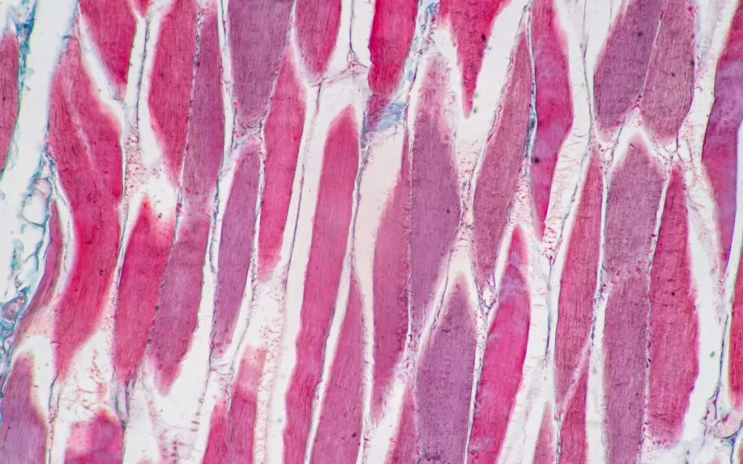
Notably, myostatin’s influence appears absent in cardiac or smooth muscle tissues. Its discovery dates back to 1997, rooted in its observed inhibitory role in muscle growth, which was elucidated through murine studies. Myostatin purportedly restrains murine satellite cell activation, representing partially committed stem cells within muscle tissue. Experimental models suggest myostatin overexpression may lead to diminished muscle mass.

Myostatin is presumed to bind with a high affinity to ActR2B receptors, initiating a signaling cascade involving Smad2/3 pivotal for muscle regulation. Other ligands within the transforming growth factor-beta (TGF-β) superfamily, such as various GDFs and activins, may bind ActR2B and regulate muscle growth. ACE-031’s mechanism appears to involve binding with circulating members of the TGF-β superfamily, particularly myostatin, potentially leaving ACV2RB receptors uninhibited[3], thereby triggering muscle hypertrophy and augmenting skeletal muscle tissue size. Additionally, ACE-031 may exert positive effects on metabolism, fat storage, and bone density.


ACE-031 Peptide and Lipid Metabolism

Investigations[4] into ACE-031’s impact on fat metabolism within the context of obesity research have unveiled intriguing insights. Recent studies have hinted at a potential correlation between myostatin expression and obesity, suggesting its involvement in metabolic pathways. Notably, in murine obesity models, elevated levels of myostatin and its receptor ActR2b have been observed compared to lean counterparts. Experimental manipulations inducing myostatin overexpression in these models have suggested a concurrent decline in muscle and myocardial mass alongside an increase in adipose tissue, implying a possible role of myostatin in promoting adiposity and reducing muscle mass.

Conversely, myostatin depletion in select murine models presents a possible research path for mitigating age-related adipose tissue accumulation and partially alleviating obesity-related traits. Furthermore, observations in mice subjected to high-calorie diets suggest that myostatin deficiency may lead to “enhanced peripheral tissue fatty acid oxidation and increased thermogenesis, culminating in increased fat utilisation and reduced adipose tissue mass.”

In light of these findings, researchers are exploring the potential of ACE-031 in obesity-related complications in murine models. Preliminary findings[5] suggest that ACE-031 may have promise in preventing and mitigating obesity-associated phenotypes.


Research in Muscle Tissue Hypertrophy

In a rigorously designed experimental framework structured as a double-blind, placebo-controlled research study,[6] the potential of ACE-031 in muscle tissue was explored alongside a comprehensive pharmacokinetic analysis. According to the pharmacokinetic analysis report, ACE-031 exhibited a half-life estimated to range between 10 to 15 days. The findings of this study suggested a potential augmentation in muscle mass following exposure to ACE-031. This inference was drawn from meticulous assessments of muscle tissue alterations, quantified through precise measurement techniques, including dual-energy X-ray absorptiometry (DEXA) and magnetic resonance imaging (MRI) conducted 29 days post-peptide exposure.

The results indicated a possible surge in muscle mass, seen in a 3.3% escalation in total lean mass and a 5.1% increase in quadriceps femoris muscle volume. Furthermore, notable shifts in serum biomarkers were observed, suggesting potential bone and fat metabolism enhancements. These percentages underscore profound alterations in muscle tissue, indicative of ACE-031’s potential to stimulate hypertrophy.


ACE-031 Peptide and Muscle Contractility

Ongoing research[7] has proposed that the peptide’s action may extend beyond myostatin inhibition. ACE-031 may enhance muscle contractile force by potentially mitigating oxidative stress within muscle tissues, conserving energy, and fostering oxidative respiration within muscles. These hypotheses stem from observations conducted in murine models, with assessments facilitated through magnetic resonance imaging (MRI) and dynamic [31P]-magnetic resonance spectroscopy ([31P]-MRS).

Specifically, exposure to ACE-031 appeared to yield a marked increase in muscle volume, with no discernible alteration in the distribution of muscle fiber types. This suggests a potential role for the peptide in promoting muscle growth. Furthermore, murine models exhibited a notable elevation in basal oxygen consumption and energy expenditure, indicative of heightened metabolic activity. During standardized fatiguing exercises, animal models exposed to ACE-031 reportedly exhibited enhanced muscle performance in a significantly higher maximum and total absolute contractile forces compared to the control group.

However, it is pertinent to note that while ACE-031 appeared to boost muscle contractile forces, it did not seem to affect the specific force-generating capacity or fatigue resistance. Moreover, metabolic fluxes, adenosine triphosphate (ATP) homeostasis, and contractile efficiency during exercise appeared to have remained largely unaffected. Intriguingly, ACE-031 seemed to diminish the intrinsic mitochondrial capacity for ATP production, hinting at potential alterations in energy generation pathways within muscle cells. However, the precise implications of this finding warrant further investigation.


Studies on Energy Metabolism

Studies have indicated that inhibiting endogenous ACE-031 proteins might not effectively lower serum lactate levels, potentially impeding the occurrence of metabolic damage to muscles and the vascularization of muscle tissue. However, supplementation with ACE-031 may potentially mitigate such impacts. ACE-031 supplementation is speculated to foster muscle cell growth by thwarting myostatin-mediated wasting. Moreover, it may postpone the onset of fatigue and oxidative damage by enhancing muscle tissue oxygenation.


ACE-031 Peptide and Bone Density

A study[8] delved into the potential action of ACE-031 on bone tissue in murine models of Duchenne Muscular Dystrophy (DMD), or muscle degeneration and heightened fracture susceptibility. The models were categorized into groups based on their activity level (running or sedentary) and further subdivided into active or placebo cohorts.

The research indicated that ACE-031 led to noticeable increases in body and muscle weights in sedentary and exercising murine models. Notably, researchers reported that femoral micro-CT analysis appeared to exhibit a substantial bone volume and trabecular number enhancement within the ACE-031 groups. While running also seemed to enhance these bone parameters in the control group, it did not notably improve trabecular bone structure or volumetric bone mineral density.

Furthermore, ACE-031 was implicated in augmenting bone mass in vertebral tissue. According to research, “the number of osteoclasts was decreased in histological analysis and the expression of several osteoblast marker genes was increased in ActRIIB-Fc treated mice, suggesting decreased bone resorption and increased bone formation in these mice.”


ACE-031 Peptide and Cancer

Molecular pathways contributing to muscle loss, often attributed to apoptosis or necrosis, are frequently observed in cancer research. The principal instigator is the metabolic strain on muscles induced by alterations in aerobic respiration status. Concurrently, an elevation in intracellular free radical levels appears to indirectly exacerbate muscle damage. Intervention with ACE-031 appears to activate the ERK1/2 pathway, thereby potentially mitigating muscle fiber atrophy stemming from apoptosis. Moreover, research indicates possible enhancements in energy utilization efficiency and mitochondrial metabolism following ACE-031 exposure, concomitant with a reduction in free radical concentration.

Furthermore, certain cancers may produce myostatin, exacerbating muscle wasting. These malignancies are often characterized by deactivated activin receptors, mitochondrial loss, and subsequently, diminished ATP levels. Studies suggest a reversal of these detrimental effects upon exposure to ACE-031. Beyond mitigating myostatin-induced muscle wasting, ACE-031 inhibition may potentially exert various ancillary downstream impacts, including enhanced insulin sensitivity, reduced fat deposition, attenuated inflammation, and improved bone metabolism and strength. Research is still ongoing.

Disclaimer: The products mentioned are not intended for human or animal consumption. Research chemicals are intended solely for laboratory experimentation and/or in-vitro testing. Bodily introduction of any sort is strictly prohibited by law. All purchases are limited to licensed researchers and/or qualified professionals. All information shared in this article is for educational purposes only.


References:

  1. National Center for Biotechnology Information (2024). PubChem Compound Summary for CID 118732224, Myostatin inhibitory peptide 7. https://pubchem.ncbi.nlm.nih.gov/compound/Myostatin-inhibitory-peptide-7.
  2. McPherron AC, Lawler AM, Lee SJ. Regulation of skeletal muscle mass in mice by a new TGF-beta superfamily member. Nature. 1997 May 1; 387(6628):83-90. https://pubmed.ncbi.nlm.nih.gov/9139826/
  3. Ozawa T, Morikawa M, Morishita Y, Ogikubo K, Itoh F, Koinuma D, Nygren PÅ, Miyazono K. Systemic administration of monovalent follistatin-like 3-Fc-fusion protein increases muscle mass in mice. iScience. 2021 May 14;24(5):102488. doi: 10.1016/j.isci.2021.102488. PMID: 34113826; PMCID: PMC8170004. https://www.ncbi.nlm.nih.gov/pmc/articles/PMC8170004/
  4. Yang M, Liu C, Jiang N, Liu Y, Luo S, Li C, Zhao H, Han Y, Chen W, Li L, Xiao L, Sun L. Myostatin: a potential therapeutic target for metabolic syndrome. Front Endocrinol (Lausanne). 2023 May 23;14:1181913. doi: 10.3389/fendo.2023.1181913. PMID: 37288303; PMCID: PMC10242177. https://www.ncbi.nlm.nih.gov/pmc/articles/PMC10242177/
  5. Zhang C, McFarlane C, Lokireddy S, Masuda S, Ge X, Gluckman PD, Sharma M, Kambadur R. Inhibition of myostatin protects against diet-induced obesity by enhancing fatty acid oxidation and promoting a brown adipose phenotype in mice. Diabetologia. 2012 Jan;55(1):183-93. doi: 10.1007/s00125-011-2304-4. Epub 2011 Sep 17. Erratum in: Diabetologia. 2015 Mar;58(3):643. PMID: 21927895. https://pubmed.ncbi.nlm.nih.gov/21927895/
  6. Attie KM, Borgstein NG, Yang Y, Condon CH, Wilson DM, Pearsall AE, Kumar R, Willins DA, Seehra JS, Sherman ML. A single ascending-dose study of muscle regulator ACE-031 in healthy volunteers. Muscle Nerve. 2013 Mar;47(3):416-23. https://pubmed.ncbi.nlm.nih.gov/23169607/
  7. Béchir N, Pecchi E, Vilmen C, Le Fur Y, Amthor H, Bernard M, Bendahan D, Giannesini B. ActRIIB blockade increases force-generating capacity and preserves energy supply in exercising mdx mouse muscle in vivo. FASEB J. 2016 Oct;30(10):3551-3562. https://pubmed.ncbi.nlm.nih.gov/27416839/
  8. Puolakkainen, Tero et al. “Treatment with soluble activin type IIB-receptor improves bone mass and strength in a mouse model of Duchenne muscular dystrophy.” BMC musculoskeletal disorders vol. 18,1 20. 19 Jan. 2017. https://www.ncbi.nlm.nih.gov/pmc/articles/PMC5244551/
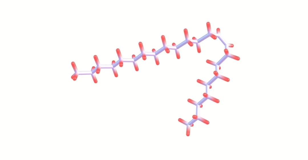
  9. Image 1 Source: https://pubchem.ncbi.nlm.nih.gov/compound/Myostatin-inhibitory-peptide-7#section=2D-Structure
AICAR Peptide: Insulin Sensitivity, Apoptosis, and Endurance

AICAR Peptide: Insulin Sensitivity, Apoptosis, and Endurance

In the realm of cellular energetics and metabolic regulation, the peptide AICAR has emerged as a pivotal research compound as it appears to offer compelling prospects for research exploration.

Analogous to adenosine monophosphate (AMP), a fundamental nucleotide pivotal in cellular energy metabolism, AICAR (short for 5-aminoimidazole-4-carboxamide ribonucleotide)[1] has garnered attention for its potential in various realms of scientific inquiry.

A significant aspect of AICAR’s potential lies in its ability to potentially reduce reperfusion injury following tissue ischemia and to potentially improve metabolic disorders. The key to how it works seems to lie in its activation of AMP-activated protein kinase (AMPK), an enzyme that plays a vital role in many metabolic processes inside cells. AMPK’s job involves bringing the cell’s energy back into balance by controlling processes that either use up energy (like making proteins and fats) or produce energy (like breaking down glucose and fats).[1] By slowing down energy-consuming processes and speeding up energy-producing ones, AMPK is considered to help cells generate more ATP, the energy currency of cells.

Moreover, the influence of AMPK extends beyond mere energy regulation, encompassing pivotal cellular processes including autophagy, mitochondrial biogenesis, and inflammatory modulation. Due to its potential to activate AMPK, AICAR peptide offers possible pathways to increase the uptake of glucose in skeletal muscle, improve sensitivity to insulin, and enhance tolerance to glucose. Additionally, scientists are considering its anti-inflammatory potential and the possibility of improving physical performance in certain experimental conditions. As such, the interplay between AICAR peptide and AMPK unveils several possibilities, requiring further investigation into its multifaceted roles.


AICAR Peptide and Organ Protection

Researchers suggest peptide may exhibit promise in conferring organ-protective impacts, particularly concerning ischemia and reperfusion injury. Initial investigations suggest that AICAR may attenuate myocardial infarction size and enhance cardiac function in an animal model subjected to myocardial ischemia-reperfusion injury.[2]

Notably, a meta-analysis encompassing data from five randomized, placebo-controlled, double-blind studies[3] further explored AICAR’s potential in cardiovascular contexts. The analysis indicated that peptide exposure may have been associated with reductions in myocardial tissue infarction size and cardiac cell death, potentially leading to improved overall outcomes. This protective potential of peptide is suggested by researchers to stem from its influence on cellular metabolism, possibly rendering cells more resilient to hypoxic conditions by upregulating energy availability, notably myocardial glucose. Experimental data from murine models suggests that AICAR, through AMPK activation, “may participate in the control of glycogen metabolism.” Furthermore, AICAR exposure was correlated with elevated levels of 5-aminoimidazole-4-carboxamide 1-beta-d-ribofuranotide (ZMP), its active intracellular form. Although AICAR did not appear to notably impact the activity of glycogen synthase (GS) or glycogen phosphorylase (GP) in tissue homogenates, it seemingly facilitated glycogenolysis through allosteric activation of GP, potentially providing an alternative energy substrate during cellular stress.[4]

Beyond its cardioprotective potential, research suggests that peptide “appears to protect the liver from fatty changes associated with chronic alcohol [exposure]” as observed in experimental murine models. Chronic ethanol exposure typically induces histological and biochemical changes indicative of fatty liver. However, AICAR intervention appeared to have attenuated these alterations, potentially by downregulating hepatic sterol regulatory element-binding protein 1c (SREBP-1c) expression and reducing fatty acid synthase (FAS) enzyme activity. SREBP-1c, a key regulator of lipid metabolism primarily in hepatic tissues, is considered by scientists to modulate the expression of genes involved in cholesterol, fatty acid, and triglyceride synthesis. Consequently, the observed decrease in SREBP-1c levels following peptide exposure likely contributes to diminished fatty acid synthesis. Meanwhile, FAS, a pivotal enzyme in fatty acid biosynthesis, appears to be regulated by SREBP-1c, further implicating AICAR in attenuating hepatic lipid accumulation.[5]


AICAR and Physical Activity

AICAR peptide has garnered considerable attention by researchers in studies in the realm of physical endurance, with researchers hypothesizing its potential to activate key metabolic pathways to improve and increase activity. Specifically, studies suggest that AICAR may activate enzymes such as AMPK, glycogen phosphorylase, and fructose-1,6-bisphosphatase, leading to potential enhancements in oxidative metabolism and the creation of new mitochondria, a process known as mitochondrial biogenesis.[6] The augmentation of mitochondrial quantity and function is suggested to confer benefits to muscle endurance. For instance, experimental data indicates that AICAR exposure in sedentary murine models appeared to have resulted in a substantial improvement in running endurance, potentially attributable to the induction of metabolic genes. These findings imply that peptides may modulate the AMPK-PPARδ pathway to facilitate training adaptations or augment endurance capacity without the need for physical exercise.[7]

PPARδ, short for Peroxisome Proliferator-Activated Receptor Delta, represents a class of nuclear receptors implicated in the regulation of genes associated with energy metabolism. It is hypothesized that PPARδ may influence processes such as lipid oxidation and mitochondrial biogenesis. The AMPK-PPARδ pathway is proposed as a conduit between the energy-sensing function of AMPK and the gene regulatory role of PPARδ. Activation of this pathway potentially induces adaptations in muscle cells akin to those induced by prolonged physical activity, including heightened mitochondrial content and a shift in muscle fiber composition towards endurance-oriented fibers, thereby potentially augmenting endurance capacity.

Further experiments in murine models have provided additional insights into the potential of peptide in augmenting endurance.[8] Notably, the introduction of an AMP-activated protein kinase agonist appeared to have resulted in increased endurance compared to controls. Additionally, in a murine model of Duchenne muscular dystrophy, AICAR appeared to have the potential to enhance the effects of physical activity and muscle function, possibly through the stimulation of autophagy.

Moreover, investigations into the vascular effects of peptide have revealed intriguing findings. Infusion of AICA-riboside, a precursor of AICAR, was associated with correlated increases in forearm blood flow, potentially mediated by nitric oxide. This suggests a potential dual role for AICAR in improving muscle blood flow and acting as a nitric oxide booster, both of which are critical factors in prolonged physical activity.[9]


AICAR Peptide and Insulin Sensitivity

Research suggests that AICAR may enhance the insulin sensitivity of various tissues by activating AMPK within cells, thereby facilitating glucose uptake. In an experimental model focusing on equine skeletal muscle, AICAR exposure appeared to lead to a decrease in glucose levels and an increase in insulin concentration, while lactate concentration remained unaffected. Notably, AICAR potentially augmented the ratio of phosphorylated to total AMPK in skeletal muscle and may have upregulated GLUT8 protein expression. The observed elevation in GLUT8 protein expression could potentially enhance glucose transport into cells, consequently improving insulin sensitivity.

Moreover, a study[10] investigating AICAR’s impact on muscle glucose uptake alongside physical activity revealed a potential increase in glucose uptake in muscle tissue. This effect might extend beyond muscle tissue, potentially enhancing peripheral and overall insulin sensitivity. Researchers also proposed that peptide might elevate the phosphorylation of extracellular signal-regulated kinase 1/2, enzymes crucial in the MAP kinase/ERK pathway, which regulates cellular processes like division, differentiation, and stress response.

Furthermore, investigations suggest that AICAR may potentially decrease hepatic glucose output, lower glucose concentrations, promote hepatic fatty acid oxidation, and inhibit lipolysis, consequently reducing plasma-free fatty acid availability.[11] Although no increase in AMPK phosphorylation was reported in skeletal muscle, a significant rise in acetyl-CoA carboxylase phosphorylation was observed. This enzyme is considered to play a pivotal role in fatty acid metabolism, catalyzing the conversion of acetyl-CoA to malonyl-CoA, a critical step in fatty acid synthesis. The apparent inactivation of acetyl-CoA carboxylase is speculated to stimulate “hepatic fatty acid oxidation and/or inhibits whole body lipolysis, thereby reducing plasma NEFA concentration.”


AICAR Peptide and Cellular Apoptosis

Data from research studies suggests that peptide may instigate programmed cell death, known as apoptosis, in test models of B-cell chronic lymphocytic leukemia (B-CLL).

Specifically, one study[12] posits that this phenomenon might entail the activation of specific enzymes involved in apoptosis, including caspase-3, -8, and -9, alongside the release of cytochrome C. Furthermore, the incubation of B-CLL cells with AICAR appears to stimulate the phosphorylation of AMP-activated protein kinase (AMPK), indicating the potential of peptide in activating this protein. Investigation into the cellular mechanisms underlying AICAR-induced apoptosis explored the necessity of AICAR’s entry into the cell and its subsequent conversion to AICA ribotide (ZMP). This inquiry employed various inhibitors, such as Nitrobenzylthioinosine (NBTI), 5-iodotubercidin, and adenosine, which were hypothesized to impede AICAR-induced apoptosis and AMPK phosphorylation. Interestingly, inhibitors targeting protein kinase A and mitogen-activated protein kinases did not seem to hinder AICAR-induced apoptosis in B-CLL cells.

Moreover, the study observed that peptide did not appear to have significantly impacted the levels or phosphorylation of p53, suggesting a mechanism of apoptosis independent of p53 activation in B-CLL cells. A comparative analysis of the sensitivity of normal B lymphocytes, T cells, and B-CLL cells to AICAR-induced apoptosis appeared to reveal similar susceptibility between normal B lymphocytes and B-CLL cells, with T cells from B-CLL subjects displaying only marginal sensitivity. Notably, the phosphorylation of AMPK was not observed in T cells exposed to AICAR. Furthermore, upon AICAR exposure, B-CLL cells appeared to have exhibited higher intracellular levels of ZMP compared to T cells, implying that the accumulation of ZMP may play a pivotal role in activating AMPK and prompting apoptosis in these cells.

Disclaimer: The products mentioned are not intended for human or animal consumption. Research chemicals are intended solely for laboratory experimentation and/or in-vitro testing. Bodily introduction of any sort is strictly prohibited by law. All purchases are limited to licensed researchers and/or qualified professionals. All information shared in this article is for educational purposes only.


References:

  1. National Center for Biotechnology Information (2024). PubChem Compound Summary for CID 65110, AICA ribonucleotide https://pubchem.ncbi.nlm.nih.gov/compound/AICA-ribonucleotide.
  2. Cieslik, K. A., Taffet, G. E., Crawford, J. R., Trial, J., Mejia Osuna, P., & Entman, M. L. (2013). AICAR-dependent AMPK activation improves scar formation in the aged heart in a murine model of reperfused myocardial infarction. Journal of molecular and cellular cardiology, 63, 26–36. https://doi.org/10.1016/j.yjmcc.2013.07.005
  3. Mangano D. T. (1997). Effects of acadesine on myocardial infarction, stroke, and death following surgery. A meta-analysis of the 5 international randomized trials. The Multicenter Study of Perioperative Ischemia (McSPI) Research Group. JAMA, 277(4), 325–332. https://doi.org/10.1001/jama.277.4.325
  4. Longnus, S. L., Wambolt, R. B., Parsons, H. L., Brownsey, R. W., & Allard, M. F. (2003). 5-Aminoimidazole-4-carboxamide 1-beta -D-ribofuranoside (AICAR) stimulates myocardial glycogenolysis by allosteric mechanisms. American journal of physiology. Regulatory, integrative and comparative physiology, 284(4), R936–R944 https://doi.org/10.1152/ajpregu.00319.2002
  5. Tomita, K., Tamiya, G., Ando, S., Kitamura, N., Koizumi, H., Kato, S., Horie, Y., Kaneko, T., Azuma, T., Nagata, H., Ishii, H., & Hibi, T. (2005). AICAR, an AMPK activator, has protective effects on alcohol-induced fatty liver in rats. Alcoholism, clinical and experimental research, 29(12 Suppl), 240S–5S. https://doi.org/10.1097/01.alc.0000191126.11479.69
  6. Hardie DG. AMP-activated protein kinase: an energy sensor that regulates all aspects of cell function. Genes Dev. 2011 Sep 15;25(18):1895-908. doi: 10.1101/gad.17420111. PMID: 21937710; PMCID: PMC3185962. https://www.ncbi.nlm.nih.gov/pmc/articles/PMC3185962/
  7. Narkar, V. A., Downes, M., Yu, R. T., Embler, E., Wang, Y. X., Banayo, E., Mihaylova, M. M., Nelson, M. C., Zou, Y., Juguilon, H., Kang, H., Shaw, R. J., & Evans, R. M. (2008). AMPK and PPARdelta agonists are exercise mimetics. Cell, 134(3), 405–415. https://doi.org/10.1016/j.cell.2008.06.051
  8. Goodyear, L. J. (2008). The exercise pill—too good to be true? New England Journal of Medicine, 359(17), 1842-1844. https://www.nejm.org/doi/abs/10.1056/NEJMcibr0806723
  9. Bosselaar, M., Boon, H., van Loon, L. J., van den Broek, P. H., Smits, P., & Tack, C. J. (2009). Intra-arterial AICA-riboside administration induces NO-dependent vasodilation in vivo in human skeletal muscle. American journal of physiology. Endocrinology and metabolism, 297(3), E759–E766. https://doi.org/10.1152/ajpendo.00141.2009
  10. Cuthbertson, D. J., Babraj, J. A., Mustard, K. J., Towler, M. C., Green, K. A., Wackerhage, H., Leese, G. P., Baar, K., Thomason-Hughes, M., Sutherland, C., Hardie, D. G., & Rennie, M. J. (2007). 5-aminoimidazole-4-carboxamide 1-beta-D-ribofuranoside acutely stimulates skeletal muscle 2-deoxyglucose uptake in healthy men. Diabetes, 56(8), 2078–2084. https://doi.org/10.2337/db06-1716
  11. Boon, H., Bosselaar, M., Praet, S. F., Blaak, E. E., Saris, W. H., Wagenmakers, A. J., McGee, S. L., Tack, C. J., Smits, P., Hargreaves, M., & van Loon, L. J. (2008). Intravenous AICAR administration reduces hepatic glucose output and inhibits whole body lipolysis in type 2 diabetic patients. Diabetologia, 51(10), 1893–1900. https://doi.org/10.1007/s00125-008-1108-7
  12. Campàs, C., Lopez, J. M., Santidrián, A. F., Barragán, M., Bellosillo, B., Colomer, D., & Gil, J. (2003). Acadesine activates AMPK and induces apoptosis in B-cell chronic lymphocytic leukemia cells but not in T lymphocytes. Blood, 101(9), 3674–3680. https://doi.org/10.1182/blood-2002-07-2339