Copper Peptide (AHK-Cu) and Hair Follicle Regeneration

Copper Peptide (AHK-Cu) and Hair Follicle Regeneration

What is AHK-Cu Peptide?

AHK-Cu is a short peptide with three amino acids attached to a copper molecule (Ala-His-Lys-Cu).  AHK-Cu peptide, also known as copper peptide, has been isolated in blood samples. This peptide may be responsible, in part, for growth and development of cells in the blood vessels. It has also been researched in relation to the generation of new blood vessels (angiogenesis). These actions of the AHK-Cu peptide are posited to be due to its potential to stimulate the production of another peptide called the Vascular Endothelial Growth Factor (VEGF). Producing new blood cells may help supply blood to biological structures such as skin and hair follicles, and may stimulate their development.

Researchers have also studied AHK-Cu in relation to its potential impact on Growth Factor Beta-1. Through its potential impact on Growth Factor Beta-1 and VEGF, AHK-Cu peptide may activate the replication and development of fibroblast cells. Fibroblasts are cells abundantly found in biological structures, including skin, hair follicles, and blood vessels. Fibroblasts are considered to activate the production of proteins such as elastin and collagen, essential participants of the extracellular matrix in the skin.


Research on AHK-Cu Peptide


Follicle Growth and Maintenance

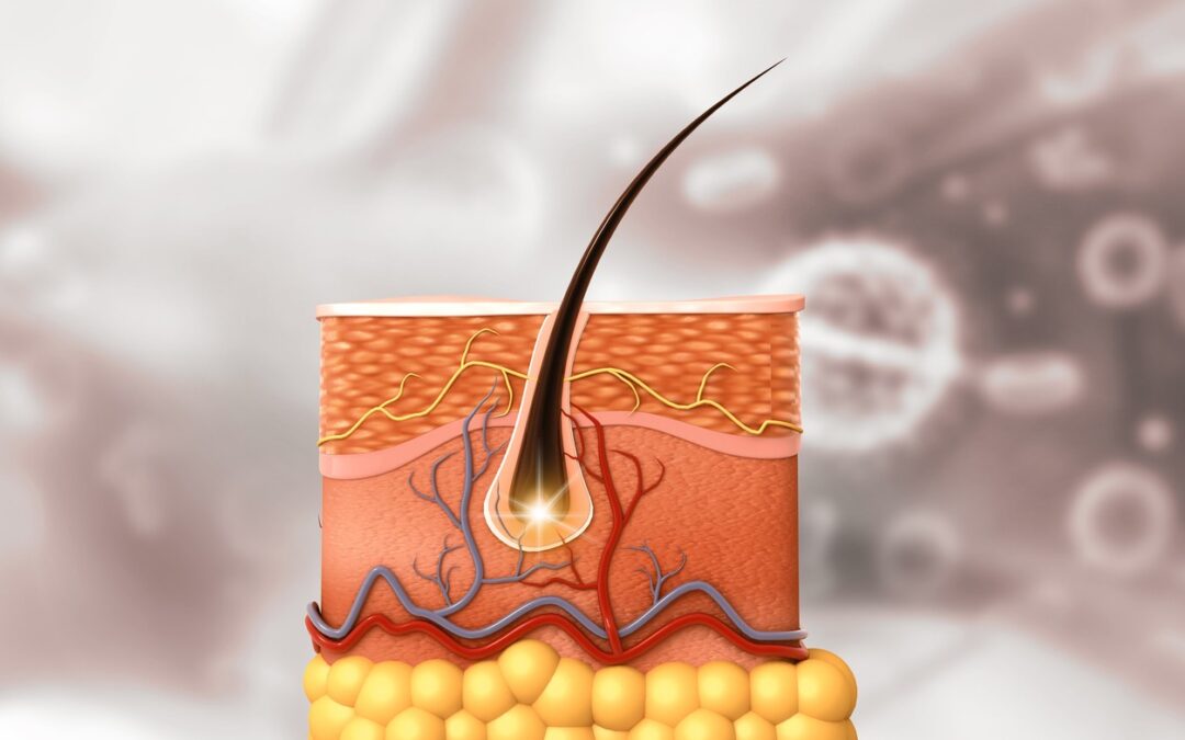
AHK-Cu peptide may potentiate immense growth on follicle development and maintenance. Researchers suggest it may supplement follicle density by preventing possible loss and by stimulating new growth. Follicles may be found in highly vascular structures, areas where extensive network of blood vessels are concentrated, to supply blood to developing follicles. The supply of blood is considered to be crucial because follicles undergo a rapid cell turnover and appear to depend on the supply of blood and nutrients in the process. Along those same lines, blood supply has been suggested to limit the production of, and stability of existing follicles. AHK-Cu peptide may potentially promote new blood vessel development by triggering the release of VEGF.

Another important phenomenon that may potentially damage follicle development is the excessive conversion of testosterone to dihydrotestosterone (DHT). Researchers consider it to be a potentially critical pathology in certain conditions characterized by immense hair loss. AHK-Cu peptide might be relevant in these contexts, as researchers suggest it may prevent the conversion of testosterone to DHT. Second, it may potentially down-regulate the action of DHT on hair follicle receptors.

The hair follicles appear to undergo three main phases of growth: anagen, catagen, and telogen phase. The anagen phase is the growth phase of follicles, and is a highly active phase. Research suggests that AHK-Cu peptide may ‘trap’ the follicles in the anagen phase of the growth cycle and maintain that phase for a considerable duration (certain research findings suggest upwards of a year).


Cell Aging and Skin Structure

A subset of skin cells, fibroblasts are deemed responsible for the secretion of skin proteins such as collagen and elastin. These proteins are considered to lend the skin structure flexibility, and suppleness. Cell aging in the skin may lead to rapid decline in the number of fibroblasts. As a result, the amount of collagen and elastin endogenously produced begins to decline as well, introducing irregularities and textural inconsistencies in the skin and developing creases along the skin surface.

AHK-Cu peptide may potentiate the reversal of cell aging by triggering the growth and development of new fibroblasts. Once there are enough fibroblasts in the extracellular matrix of the skin, natural elastin and collagen production may follow. Research suggests that AHK-Cu may potentially stimulate the production of Type 1 collagen by up to 300%.


Disclaimer: The products mentioned are not intended for human or animal consumption. Research chemicals are intended solely for laboratory experimentation and/or in-vitro testing.  Bodily introduction of any sort is strictly prohibited by law.  All purchases are limited to licensed researchers and/or qualified professionals. All information shared in this article is for educational purposes only.


Comparing Ipamorelin and GHRP-6

Comparing Ipamorelin and GHRP-6

Growth Hormone-Releasing Peptides (GHRPs)

Growth hormone-releasing peptides GHRPs are classified as synthetic peptides capable of inducing stimulatory effects on the endogenous secretion of growth hormone (GH). Research studies conducted on animal models have led scientists to explore and classify several research peptides within this family of peptides. However, researchers note that they appear to have no structural resemblance with Growth Hormone-Releasing Hormones and may potentially mediate their action by binding with specific cognate receptors present at the pituitary or hypothalamic level.


Ipamorelin

Ipamorelin is considered to be one of the most versatile synthetic peptides classified as a GHRP. Composed of only five amino acids, it appears to be a growth hormone secretagogue receptor (GHS-R) agonist. Research studies have posited that it may be relevant in various branches of scientific research, and may be implicated in gastrointestinal functioning, growth hormone production, and neurological impacts. Ipamorelin is considered by researchers to be a ‘gentle peptide’ compared to other peptides like Sermorelin or GHRP-6, as they assert it appears to affect only the growth hormone axis.

The binding of Ipamorelin peptide has been suggested to occur upon the ghrelin (growth hormone secretagogue) receptor. The ghrelin receptor (a.k.a. GHS-R) in the brain, liver, heart, and muscular structures, may potentiate the regulation of energy homeostasis and certain metabolic processes. It appears to be highly exclusive for growth hormone release from the pituitary gland.

Both Ipamorelin and a similar agonist, GHRP-6, belong to the class of third-generation GHRP. They are among the first synthetic peptides studied extensively in animal models. Ipamorelin and GHRP-6 appear to be similar in their functions. Both peptides, for example, have been suggested to release growth hormone at similar rates. However, there appears to be a moderate difference in how the two peptides mediate their action. GHRP-6 may potentially cause a release and an increase in cortisol and prolactin levels, whereas Ipamorelin potentially only selectively releases growth hormone.


Growth Hormone-Releasing Peptide-6

Growth hormone-releasing peptide-6 is a first-line synthetic hexapeptide and a met-enkephalin derivative. It comprises a chain of 6 synthetic D amino acids with the sequence (His-D-Trp-Ala-Trp-D-Phe-Lys-NH2). Additionally, the peptide also contains two methyl groups as modifications. It happens to be the first hexapeptide studied in animal model studies, and researchers identified it more than a decade ago.

GHRP-6 has been suggested by researchers to recognize and bind to specific receptors present at the pituitary or hypothalamic glands. Studies have suggested that GHRP-6 receptors are abundant in different peripheral tissues such as the heart, adrenal, ovary, testis, lung, and skeletal muscle.
Studies in animal test models posit that GHRP-6 may stimulate ghrelin response. Ghrelin, in turn, may increase the production of growth hormone (GH) by acting at the level of the pituitary or hypothalamus through a specific receptor that is different from that of the endogenous Growth Hormone-Releasing Hormone (GHRH). The four essential organs of the test subjects that have been observed to be influenced under exposure to GHRP-6 include the pituitary gland, central nervous system, liver, and stomach.

Research studies in animal models have highlighted that GHRP-6 and Ipamorelin may potentially stimulate ghrelin, “the hunger hormone,” by associating with its ghrelin receptor. This association may trigger a cascade of events by inducing increased growth hormone production.

Іn comparison with GHRP-6, Iраmоrеlіn appears to induce a more selective activation of GHRH as it does not appear to have any effect on cortisol or prolactin. It may potentially metabolize slower than GHRP-6, and may possibly remain present in the bloodstream for a longer duration, adding to its stability.


Disclaimer: The products mentioned are not intended for human or animal consumption. Research chemicals are intended solely for laboratory experimentation and/or in-vitro testing.  Bodily introduction of any sort is strictly prohibited by law.  All purchases are limited to licensed researchers and/or qualified professionals. All information shared in this article is for educational purposes only.
Thyrotropin and Research Regarding Metabolism

Thyrotropin and Research Regarding Metabolism

Thyrotropin, also recognized as thyroid-stimulating hormone (TSH) or thyrotropic hormone, are naturally secreted by the thyrotropic cells of the adenohypophysis. It is classified as a glycoprotein hormone that researchers propose regulates the endocrine activity of the thyroid gland. It may bind to specific receptors on the surface of the thyroid glands, potentially leading to the cleavage of thyroglobulin (a large protein stored in the follicles within the thyroid gland) and the potential production of Thyroxine T4 (Tetraiodothyronine) and Triiodothyronine (T3).

The speculation is that Thyrotropin might influence the synthesis of thyroglobulin and the growth of thyroid cells. Researchers suggest that the paraventricular cells of the hypothalamus might secrete a Thyrotropin-releasing hormone (TRH), which then may command the release of the thyrotropin hormone. This release might form a hypothalamic pituitary thyroid axis in which the regulation of the secretion of hormones could occur through a feedback mechanism. It is suggested that whenever there is excess thyroxine in the circulation, this might act on the specific receptors of the pituitary, potentially suppressing the level of thyrotropin and, ultimately, Thyrotropin-Releasing Hormone. If there is insufficient thyroxine in the circulation, researchers propose that the positive feedback from elevated thyrotropin levels may stimulate the release of further thyroxine.

Thyrotropin levels might naturally be elevated in primary hypothyroidism and suppressed in thyrotoxicosis.The circulating levels of thyrotropin may manifest circadian and pulsatile variation, with concentration exceptionally high during rapid growth and development and increased metabolic demand. Another hormone released by the hypothalamus, called Somatostatin, might antagonize the action of Thyrotropin.

Thyrotropin is considered a glycoprotein consisting of Alpha and Beta subunits. The alpha subunit appears to be similar to other glycoprotein hormones such as Luteinizing hormone (LH), Follicle Stimulating Hormone (FSH), and Chorionic Gonadotropin (HCG). Conversely, the beta chain might differ in these glycoproteins, imparting unique biological characteristics. Researchers suggest that these glycoproteins function through Cyclic Adenosine Monophosphate (cAMP), potentially signaling the second messenger system. The dual messenger system may convert Adenosine monophosphate into Cyclic adenosine monophosphate.

Thyrotropin might activate another signaling cascade, called the Inositol Triphosphate second messenger system (IP3). Researchers propose that Thyrotropin might activate specific G protein-coupled receptors on the surface of thyroid follicular cells, leading to the activation of both cAMP and IP3 /Ca+2 pathways. This activation might ultimately enhance iodine uptake in the follicular thyroid cells, and an array of further steps might lead to the formation of T4 and T3, considered to be responsible for all the downstream physiological effects of the thyroid hormone.


Research Implications of Thyrotropin

Thyrotropin has been hypothesized to impact various physiological processes via the production of Thyroxine T4 and Triiodothyronine T3.

Regulation of metabolism: Thyrotropin peptide might stimulate the thyroid gland’s secretion of Thyroxine T4 and Triiodothyronine T3. This hormone may activate various metabolic processes, including increased glucose absorption, gluconeogenesis, glycogenolysis, lipolysis, and synthesis/degradation of the protein.

  • All of these phenomena could lead to a net state of catabolism.
  • The speculation is that the thyroid hormone might regulate metabolic rate, and thereby weight.
  • It may help maintain temperature homeostasis in animal models.
  • It may regulate the rate of the regeneration process of various tissues.
  • The thyroid hormone might affect gastric emptying.
  • It may control muscle contraction.

Potential of Thyrotropin as a Screening Test: Speculatively, it might be the first-line screening test in diagnosing both a state of hypothyroidism and hyperthyroidism. Measuring the thyrotropin level may prove to be a better method than measuring direct T3 and T4. This consideration was formed for two reasons. First, the thyrotropin levels may be affected before the T3 and T4 groups appear to change in any pathological state. Secondly, it may be more reliable than T3 and T4 levels as these fluctuate and remain volatile.


Disclaimer: The products mentioned are not intended for human or animal consumption. Research chemicals are intended solely for laboratory experimentation and/or in-vitro testing.  Bodily introduction of any sort is strictly prohibited by law.  All purchases are limited to licensed researchers and/or qualified professionals. All information shared in this article is for educational purposes only.
Insulin-like Growth Factor 1 (IGF-1)

Insulin-like Growth Factor 1 (IGF-1)

IGF-1 (Insulin-like growth factor 1), also known as somatomedin C, shares structural similarities with insulin. It is a hormone that has been suggested to play a role in the growth of developing organisms and may have anabolic effects on mature subjects, as reported via research findings in various animal studies. It may synthesize as an endocrine hormone and, as researchers propose, target organs as a paracrine or autocrine hormone. It is speculated to be produced throughout a lifetime and may be present in its highest levels during adolescence and lowest just following birth.


The Structure of IGF-1

The peptide consists of 70 amino acids with a molecular weight of 7649 Da. The disulfide bonds of IGF-1 may link the alpha and beta chains. Rat IGF-1 is suggested to synthesize as four precursor isoforms with alternating N-terminal and C-terminal propeptides. Mature IGF-1 is speculated to be identical between isoforms, and cleavage of the propeptide terminal is considered to produce it. There are 12 amino acids in the C-peptide region.

Mechanism of action studies on laboratory animals have suggested that Insulin-like Growth Factor 1 may be a significant mediator of growth hormone action. The pituitary gland is considered to produce growth hormone (GH), which circulates in the bloodstream, delivering signals to the liver to produce IGF-1.

Insulin-like Growth Factor 1 is speculated to bind to at least two of the receptors of cell membrane tyrosine kinases, including the IGF-1 receptor and the insulin receptor. Its main action, researchers suggest, is binding to the IGF-1 receptor, which may be ubiquitous in tissues. This ligand-receptor complex might stimulate cell growth and proliferation and emit intracellular messages via the AKT pathway known to inhibit programmed cell death. Insulin may also potentially bind to the IGF-1 receptor. Still, its affinity is suggested to be much lower, and Insulin-like Growth Factor 1 may also bind to the insulin receptor to produce a 10% effect on insulin’s potency. Researchers propose that Insulin-like Growth Factor 1 may interact with all seven IGF-1 binding proteins, specifically IGFBP2 and IGFBP5, and these two serum levels might be inversely proportional to circulating IGF-1.

Blood-transported IGF-1 has been speculated by research teams to regulate balanced growth across many tissues and organs. Stimulation of autocrine or paracrine IGF-1 might cause excessive growth as it works unaffected by growth hormones.


Insulin-like Growth Factor-1 Potential Effects

The first recorded action of the growth-stimulating effect of extrinsic IGF-1, researchers suggest, occurred by exposing a purified hormone to hypophysectomy rats.

  • In diabetic rats, it may be suggested to help control glucose, and efficacy might be inversely proportional to the duration of diabetes.
  • Exposure to IGF-1 may exhibit anabolic effects with subsequent rapid growth in neonatal rats, but there may be evidence that complete expression at a given concentration requires nutrition.
  • Exposing animals to Insulin-like Growth Factor 1 might induce hepatoprotective and antifibrinolytic effects in experimental cirrhosis. These effects could be associated with decreased liver levels of several factors involved in oxidative damage, such as myeloperoxidase and nitric oxide.
  • Obstruction of the right middle cerebral artery, researchers propose, induces ischemia, and IGF-1 expression in the central nervous system may reduce significantly. The potential exposure to Insulin-like Growth Factor 1 could increase expression in the affected muscles, sciatic nerve, lumbar spinal cord, and motor cortex. This exposure might reduce neuronal apoptosis and improve motor function.
  • Immunohistochemical analysis of rat testes of various ages might reveal increased IGF-1 receptors from birth to 20 days after delivery. Tests suggest that IGF reduces Leydig cell apoptosis at all stages of development.


Disclaimer: The products mentioned are not intended for human or animal consumption. Research chemicals are intended solely for laboratory experimentation and/or in-vitro testing.  Bodily introduction of any sort is strictly prohibited by law.  All purchases are limited to licensed researchers and/or qualified professionals. All information shared in this article is for educational purposes only.
The Antifibrotic Potential of B7-33 Peptide

The Antifibrotic Potential of B7-33 Peptide

B7-33 is a single-chain peptide derived from a naturally occurring compound known as H2-Relaxin. Relaxin proteins, including Relaxin, Insulin-like peptide 3, h2-relaxin, and insulin-like peptide 5, are suggested to form a family of four proteins with pleiotropic actions affecting the cardiovascular, musculoskeletal, and reproduction systems. These actions may be mediated through four types of receptors, namely RXFP1/2 and RXFP3/4. Various ligands, such as cAMP and corticotropin-releasing hormone, are proposed to stimulate these receptors, expressing antioxidant, anti-inflammatory, and wound-healing characteristics. Additionally, Relaxin is speculated to exhibit antihypertrophic, vasodilator, and angiogenic characteristics.


B7-33 Specifications:

Sequence: VIKLSGRELVRAQIAISGMSTWSKRSL

PubChem SID: 318164840

Synonyms: (B7-33) H2, GTPL9321


Mechanism of Action:

Researchers suggest that B7-33 potentially exceeds the capability of the Relaxin protein to preferentially act through the pERK Pathway rather than the cAMP-mediated pathway. Additionally, it may have a greater affinity to bind to RXFP1 receptors than Relaxin. However, the antifibrotic potential of the peptide is suggested to be mediated through RXFP1-angiotensin II type 2 receptors stimulation. It might further activate the pERK1/2 pathway, potentially leading to enhanced production of Matrix Metalloproteinase (MMP)-2. These collagen-degrading proteins could play a role in controlling fibrosis and preventing scarring. This potential of B7-33 may be induced through pERK1/2 without activating the cAMP pathway. Researchers suggest this is important because studies indicate cAMP to have a tumor-promoting potential, the most dreadful impact associated with the full-sequence Relaxin. Another speculated advantage of B7-33 over H2-relaxin protein is its less complicated structure. It may make it less laborious to produce it in the laboratories while retaining all the properties associated with the native Relaxin proteins.


Research Implications of B7-33:

  1. Antifibrotic Potential: Fibrosis (scarring) occurs following significant tissue damage, as the cells knit back together in an irregular consistency to the surrounding tissues. Researchers suggest internal fibrosis may be a significant factor in chronic inflammatory diseases. Chronic liver, heart, or lung disease fibrosis is speculated to be the leading cause of organ failure. It has been suggested that controlling this unorganized tissue regeneration may prevent organ failure. A study on H2-relaxin proteins suggest their potential to reduce fibrosis following an ischemic injury to the heart. The peptide appeared to induce an immediate vasodilatory effect in the heart that might lead to a speculated reduction in long-term scarring.  A study performed on rat models suggests that exposure to B7-33 reduced scarring by approximately 50% following significant tissue damage. This speculated reduction in fibrosis might eventually lead to improved cardiac function with lesser long-term complications associated with heart failure.
  2. Blood Vessel Protection and Preeclampsia: Researchers suggest that B7-33 possesses vasoprotective potential, comparative to Relaxin-2 (Serelaxin) against long-term scarring and endothelial dysfunction. It appears to do this through the activation of bradykinin-mediated relaxation of arteries that is endothelium-dependent. B7-33 might be more selective in its action as compared to Serelaxin.

Preeclampsia is considered to be a common complication of pregnancy that may prove life-threatening to the mother and the fetus. It is characterized in part by high blood pressure in the mother and reduced fetal weight. A recent study provided data to suggest that B7-33, by stimulating RXFP-1 receptors, may lead to enhanced Vascular Endothelial Growth Factor (VEGF) production. VEGF might stimulate the production of the cytotrophoblast cells in the fetus, which are speculated to be responsible for developing blood flow from mother to fetus. B7-33 might help improve the fetus’s survival by prolonging the speculated duration of pregnancy in cases of premature delivery.


Disclaimer: The products mentioned are not intended for human or animal consumption. Research chemicals are intended solely for laboratory experimentation and/or in-vitro testing.  Bodily introduction of any sort is strictly prohibited by law.  All purchases are limited to licensed researchers and/or qualified professionals. All information shared in this article is for educational purposes only.

Humanin and Cardiovascular Research

Humanin and Cardiovascular Research

Humanin is a micro-peptide featuring a three-turn alpha-helix and an asymmetrical structure. The number of amino acids in the peptide chain is suggested to depend on the peptide site, with 21 amino acids produced in mitochondria and 24 amino acids made in the cell’s cytosol.

Mitochondria are considered to play complex roles in energy production, cellular signaling, and metabolism, serving as the cell’s powerhouse. Earlier assumptions about the control of mitochondria’s function, maintenance, and biogenesis by the nucleus may be reconsidered in light of newer studies suggesting that some mitochondria possess a genomic sequence, translating into proteins that may affect the genomic expression of the nucleus.

Research on the genomic sequence of mitochondria suggests that the rRNA possesses Open Reading Frames (ORFs), transcribing into small peptides, including Humanin (HN), Small Humanin-Like Peptides 1-6 (SHLPs), and Mitochondrial ORF-encoded peptide (MOTS-c). These peptides appear to bind to specific receptors in and out of the cells, exerting various speculated biological effects.

The detectable concentration of Humanin in the skeletal muscles, hypothalamus, and cortex of murine models declined progressively with advancing age. Humanin may potentially be a subsequent biomarker of age. Humanin is suggested to have diverse biological functions, including cytoprotective and metabolic roles. Cytoprotective roles may involve protection against oxidative stress and injury, anti-inflammatory responses, neuroprotection, and cardioprotection. Metabolic protection roles may involve metabolic hemostasis, ATP production, reduction in visceral fat, and glucose-stimulated insulin secretion.

Myocardial fibrosis may appear either as an age-related physiological change or due to various pathological phenomena such as Myocardial Infarction and ischemic reperfusion injury, contributing to cardiac dysfunction. It is speculated to affect both systolic and diastolic function and eventually progress to heart failure. The number of fibroblast cells present in cardiac tissue may directly correlate with the age of the heart tissue. Considering its ratio to the cardiac striated cells present may indicate the extent of age-related degenerative change in the heart. These fibroblasts may lead to the secretion of extracellular matrix proteins such as collagen type 1 that favors fibrosis.


Humanin – Mechanism of Action

Studies suggest that Humanin may exert cardioprotective effects by activating the AKT/GSK-3 beta (Glycogen Synthase Kinase) pathway. Humanin might also be able to alter the pro-apoptotic factors in the cardiac fibers and prevent cell death, starting and downregulating varying pathways depending on the action site and the pathology type. Other factors considered to be responsible for causing fibrosis in the heart, such as FGF-2 (Fibroblast Growth Factor-2), MMP-2 (Matrix Metalloproteinase-2), and Transforming Growth Factor-beta (TGF-beta), may increase in the cardiac tissue as age progresses. Studies suggest the role of Humanin in attenuating the effect of them all.

Another speculated mechanism associated with age-related cardiac tissue deterioration involves the damage caused by Reactive Oxidative Species (ROS). Excessive Reactive Oxidative Species may cause the deterioration of the Antioxidant Defense System by depolarizing the mitochondrial membrane. This could lead to ATP (Adenosine Triphosphate) hydrolysis and the speculated swelling of the mitochondria. This might cause the rupture of the outer membrane of mitochondria and, eventually, the release of pro-apoptotic proteins in the cytosol. The Humanin peptide may induce enzymes and various small non-enzymatic molecules that lead to an overall reduction of the oxidative stress caused by Reactive Oxygen Species (ROS).

A study on aged rats propose that chronic exposure to exogenous Humanin may ameliorate myocardial fibrosis and apoptosis. This study indicate an increase in the ratio of cardiac fibers to fibroblasts, measured in the percentage of each cell present in a random field chosen after immunofluorescence staining of the cardiac tissue. Picrosirius red staining of the cardiac tissue might inducate a decrease in the collagen content of the heart tissue, a marker of tissue age. Long-term supplementation of Humanin may reduce fibroblast proliferation and downregulate the expression of Fibroblast Growth Factor-2 (FGF-2), Matrix Metalloproteinase-2 (MMP-2), and Transforming Growth Factor-beta 1 (TGF-beta 1). It has also been speculated to suppress pro-apoptotic factors in the cardiac tissue.


Disclaimer: The products mentioned are not intended for human or animal consumption. Research chemicals are intended solely for laboratory experimentation and/or in-vitro testing.  Bodily introduction of any sort is strictly prohibited by law.  All purchases are limited to licensed researchers and/or qualified professionals. All information shared in this article is for educational purposes only.