The
CJC-1295 & GHRP-6 blend is believed to work synergistically to stimulate the increase of growth hormone (GH). There is speculation that the increase in GH levels due to this blend might be higher than using either of these peptides alone, as well as initiate downstream action, as in the case of GHRP-6, which is believed to potentially impact hunger hormone signaling and sleep cycle regulation.
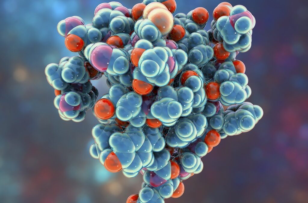
CJC-1295 Peptide
CJC-1295 is a synthetic derivative of the naturally occurring growth hormone-releasing hormone (GHRH). It has a modified sequence of the first 29 amino acids in the GHRH that is part of the growth hormone secretagogues group. This group is believed to act on the anterior pituitary and potentially causes growth hormone release. CJC-1295 is speculated to be somewhat similar to Sermorelin but varies from other growth hormone secretagogues. An additional compound called the drug affinity complex, or DAC, is believed to attach to the structure of the peptides via a lysine linker. This complex is speculated to protect the peptide from hydrolysis during its circulation in the bloodstream and, at the same time, promotes its binding to albumin, allowing it to have a potentially longer and more stable half-life in comparison to other such compounds.
CJC-1295 and Growth Hormone Release: Since CJC-1295 is classified as a growth hormone secretagogue, it might not come as a surprise that it is speculated to cause the potential stimulation of growth hormone release from the anterior pituitary. Studies conducted on lab rats are believed to show that the introduction of CJC-1295 might boost growth hormone by up to 2x – 10x, causing a peak in the growth hormone concentration after two hours of exposure, which researchers report is maintained for 6 to 10 days.
However, one key factor differentiating CJC-1295 from other growth hormone secretagogues is that it appears to affect the hormone release purely physiologically. Exposure to the peptide is believed to allow the achievement of the usual peaks and valleys in growth hormone release during the normal circadian rhythm. Meanwhile, it is also believed to block the rise in the growth hormone level to the extent that it might cause unintended downstream action. Therefore, CJC-1295 is reputed for its potential physiological effect on growth hormone release, which might help benefit from its functions without worrying about its side effects.
It has been observed in research studies, that CJC-1295 exposure may positively influence all of the effects of growth hormones, such as protein synthesis, fat metabolism, blood sugar regulation, growth (via hypertrophy and hyperplasia), preservation of myocardial function, and bone density.
Insights on CJC-1295 and Growth Studies on mice with GHRH deficiency suggest that CJC-1295 exposure is speculated to mimic the normal physiology of GHRH release. CJC-1295 is believed to cause a pulsatile increase in GH levels. This makes it an eligible candidate for research into growth hormone deficiency and related disorders.
CJC-1295 and Infertility: Research studies from the early 1990s have hinted that CJC-1295 might help mitigate the percentage of infertility in female species. This speculation is believed to be due to IGF-1 regulating ovulation. CJC-1295 is also believed to regulate the IGF-1 via a stimulation of growth hormone release, and researchers may test it in a laboratory setting for ovulation induction and even sperm production and maturation.
GHRP-6 Peptide
GHRP-6 peptide is a growth hormone secretagogue that appears to bind to ghrelin receptors and thereby induce growth hormone release by activating these receptors. This peptide consists of a sequence of 6 amino acids in its structure.
GHRP-6 and Memory: GHRP-6 is believed to improve cognitive function and memory recall by increasing physical activity. Although this mechanism of action is not quite well understood, there is speculation that there is a reason to believe that exercise and physical activity improves learning and memory, especially if it is done following a task. Research on laboratory rats suggests that GHRP exposure solidified long-term memory and converted short-term memories into long-term ones by improving blood flow to different brain regions.
Protection of Brain Tissue: Significant data suggests that GHRP-6 exposure might protect neurons and other nervous tissues by inactivating apoptotic pathways and improving blood flow, thus preventing ischemic death.
Scar Formation: Research indicates that GHRP-6 exposure might improve the appearance of the skin and reduce scarring by increasing the deposition of extracellular matrix proteins and collagen, thereby improving scar formation.
Disclaimer: The products mentioned are not intended for human or animal consumption. Research chemicals are intended solely for laboratory experimentation and/or in-vitro testing. Bodily introduction of any sort is strictly prohibited by law. All purchases are limited to licensed researchers and/or qualified professionals. All information shared in this article is for educational purposes only.