Potential Research Applications of PE-22-28 Peptide

Potential Research Applications of PE-22-28 Peptide

PE-22-28 is a synthetic peptide analog of spadin, a protein naturally produced in the central nervous system. The sequence of PE-22-28 is based on a fragment of the spadin protein that corresponds to the chain from the 22nd to 28th amino acids of its sequence. More specifically, the amino acid sequence of PE-22-28 is Proline – Glutamine – Glycine – Aspartic acid – Asparagine – Cysteine – Cysteine, with the two cysteine residues forming a disulfide bond that may contribute to the overall stability of the molecule.
The peptide was first synthesized in 2017 by Djillani et al. as a short but potentially potent analog of spadin.[1] Similar to spadin, PE-22-28 peptideappears to interact with various ion channels in the central nervous system, particularly the two-pore domain potassium channel TREK-1 (TWIK-related K+ channel 1). TREK-1 is also found in the pancreas, lungs, kidneys, heart, and muscle tissues.

Scientists consider TREK-1 to be widely expressed in the central nervous system,  playing a possible role in pain perception, mood regulation, and neuroprotection. PE-22-28 has exhibited potential binding activity to the extracellular domain of TREK-1, thereby inhibiting the channel’s activity. Further research is needed to fully understand the mechanisms of action and potential research applications of PE-22-28 peptide. PE 22–28 has also been used as the core peptide to design other analogs with different N- and C-terminal end modifications. 


PE-22-28 Peptide and TREK-1 Inhibition

The primary potential  mechanism of action of PE-22–28 peptide is considered by researchers to be exerted via blocking the TREK-1 channels in the central nervous system. According to Djillani et al., PE-22–28 is “the shortest, most efficient sequence capable of blocking the TREK-1 channel with higher potency” than spadin.[2] TREK-1 is generally the most studied background two-pore domain potassium channel. Its main role is considered to be controling cell excitability and maintaining the membrane potential below the threshold of depolarization.[3] TREK-1 may also be multi-regulated by a variety of physical and chemical stimuli. TREK-1 has also been reported in the heart and several other organs, but inhibition of these channels has unknown impact.[4]

By blocking TREK-1, murine studies suggest that PE-22–28 may significantly decrease immobility time in the forced swim test (FST) compared to control mice that received saline.[1] The FST is a common test used to measure depressive behavior in rodents, where the mice are placed in a water tank, and their mobility is monitored. In addition, the study also evaluated the potential of PE-22–28 on the learned helplessness test (LHT), another test with similar indications as FST. The LHT measures the time it takes for mice to escape from an aversive stimulus. The study commented that the sub-chronic presentation of PE-22–28 appears to significantly reduce the escape latencies in the LHT.

Furthermore, the trial evaluated the potential of PE-22-28 on a mice model of long-term corticosterone presentation.[1] Corticosterone is a hormone that is involved in the body’s response to stress, and chronic exposure to high levels of corticosterone has been associated with depressive behavior in rodents. The results of this study suggested that PE-22–28 peptide may result in decreasing immobility time in the FST. It may also reduce the latency to eat in the novelty-suppressed feeding (NSF) test, which measures an animal’s willingness to eat in a stressful environment.


PE-22-28 Peptide and Serotonin Transmission

Researchers posit that PE-22–28 may inhibit TREK-1, possibly leading to the excitation of the dorsal raphé nucleus and the firing of serotonin transmission.[5] The researchers suggest that “if a viral vector that leads to the secretion of PE 22-28 is [presented to] the dorsal raphé nucleus, the peptide will block the channel and thereby activate the serotonergic neurons, resulting in the facilitation of serotonergic transmission, as SSRIs do.

Studies suggest that PE-22–28 peptide may act as a blocker of the TREK-1 channel, similar to spadin, and Maati et al. investigated these actions in relation to the connectivity between the medial prefrontal cortex (mPFC) and dorsal raphé serotonergic neurons.[6] The study commented that spadin might increase the firing rate of serotonin neurons and that the action of spadin and 5-HT4 agonists appeared additive and independent of each other. However, adding a mGluR2/3 antagonist reportedly blocked the action of spadin, suggesting that spadin likely depends on mPFC TREK-1 channels coupled to mGluR2/3 receptors. Therefore, the researchers further suggested that PE-22-28 may also interact with the mGluR2/3 receptors, similar to spadin, to fire up the serotonin neurons. 

The mGluR2/3 is a metabotropic glutamate receptor found in the central nervous system. They are G protein-coupled receptors that are considered to be activated by the neurotransmitter glutamate. There are two subtypes of mGluR2/3 receptors: mGluR2 and mGluR3, and they are both involved in regulating neurotransmitter release.[7] Activation of mGluR2/3 receptors appears to inhibit the release of glutamate and other neurotransmitters, such as GABA and dopamine. This mechanism may help to regulate synaptic transmission and maintain the balance of excitatory and inhibitory neurotransmission in the brain. The study also suggested that the combination of 5-HT activators should be cautiously approached.[6]


PE-22-28 Peptide and Neuroplasticity

One in vitro trial suggested that inhibiting TREK-1 may increase neuronal membrane potential and activate both MAPK and PI3K signaling pathways in a time- and concentration-dependent manner.[8] The latter pathway has been linked to the supposed protective action of spadin against apoptosis. Inhibiting TREK-1 also appeared to enhance mRNA expression and protein levels of two markers of synaptogenesis, PSD-95, and synapsin. Inhibiting TREK-1 may increase the proportion of mature spines in cortical neurons, suggesting increased neuroplasticity. Inhibiting TREK-1 appeared in other experimental models to increase mRNA expression and protein levels of brain-derived neurotrophic factor (BDNF) in the hippocampus, suggesting a neuroplasticity potential.[8]

In one murine model, PE-22-28 was presented for 4 days, and on the 5th day, the brains of the rats were analyzed.[1] The results suggested that PE-22-28 significantly increased the number of bromodeoxyuridine (BrdU) positive cells in the hippocampus compared with saline mice, suggesting that PE-22-28 may induce hippocampal neurogenesis similar to spadin. BrdU is a thymidine analog that incorporates the DNA of dividing cells during the S-phase of the cell cycle.


Conclusion

PE-22-28 is a synthetic peptide analogous to spadin, a protein naturally produced in the central nervous system and considered by researchers to possibly inhibit the action of TREK-1. As a result, the peptide appears to upregulate serotonin production, possibly improving neuroplasticity and reducing depressive behavior in animal models. Nevertheless, more research is needed to evaluate its potential actions.

Disclaimer: The products mentioned are not intended for human or animal consumption. Research chemicals are intended solely for laboratory experimentation and/or in-vitro testing.  Bodily introduction of any sort is strictly prohibited by law.  All purchases are limited to licensed researchers and/or qualified professionals. All information shared in this article is for educational purposes only.


References


    1. Djillani, A., Pietri, M., Moreno, S., Heurteaux, C., Mazella, J., & Borsotto, M. (2017). Shortened Spadin Analogs Display Better TREK-1 Inhibition, In Vivo Stability and Antidepressant Activity. Frontiers in pharmacology, 8, 643. https://doi.org/10.3389/fphar.2017.00643
    2. Djillani, A., Pietri, M., Mazella, J., Heurteaux, C., & Borsotto, M. (2019). Fighting against depression with TREK-1 blockers: Past and future. A focus on spadin. Pharmacology & therapeutics, 194, 185–198. https://doi.org/10.1016/j.pharmthera.2018.10.003
    3. Djillani, A., Mazella, J., Heurteaux, C., & Borsotto, M. (2019). Role of TREK-1 in Health and Disease, Focus on the Central Nervous System. Frontiers in pharmacology, 10, 379. https://doi.org/10.3389/fphar.2019.00379
    4. Fink M, Duprat F, Lesage F, Reyes R, Romey G, Heurteaux C, Lazdunski M. Cloning, functional expression and brain localization of a novel unconventional outward rectifier K+ channel. EMBO J. 1996 Dec 16;15(24):6854-62. PMID: 9003761; PMCID: PMC452511.
    5. Okada, M., & Ortiz, E. (2022). Viral vector-mediated expressions of venom peptides as novel gene therapy for anxiety and depression. Medical Hypotheses, 166, 110910.
    6. Moha ou Maati, H., Bourcier-Lucas, C., Veyssiere, J., Kanzari, A., Heurteaux, C., Borsotto, M., Haddjeri, N., & Lucas, G. (2016). The peptidic antidepressant spadin interacts with prefrontal 5-HT(4) and mGluR(2) receptors in the control of serotonergic function. Brain structure & function, 221(1), 21–37. https://doi.org/10.1007/s00429-014-0890-x
    7. Jin LE, Wang M, Galvin VC, Lightbourne TC, Conn PJ, Arnsten AFT, Paspalas CD. mGluR2 versus mGluR3 Metabotropic Glutamate Receptors in Primate Dorsolateral Prefrontal Cortex: Postsynaptic mGluR3 Strengthen Working Memory Networks. Cereb Cortex. 2018 Mar 1;28(3):974-987. doi: 10.1093/cercor/bhx005. PMID: 28108498; PMCID: PMC5974790.
    8. Devader, C., Khayachi, A., Veyssière, J., Moha Ou Maati, H., Roulot, M., Moreno, S., Borsotto, M., Martin, S., Heurteaux, C., & Mazella, J. (2015). In vitro and in vivo regulation of synaptogenesis by the novel antidepressant spadin. British journal of pharmacology, 172(10), 2604–2617. https://doi.org/10.1111/bph.13083
Cell Proliferation and Gene Expression in Regards to Vilon Peptide

Cell Proliferation and Gene Expression in Regards to Vilon Peptide

Vilon peptide was developed in the 1990s by researchers at the Institute of Immunology in Moscow, Russia. The development of Vilon was part of a broader research program aimed at developing novel immunomodulatory peptides. The researchers were particularly interested in peptides that could regulate the activity of T-cells, a type of white blood cell that plays a key role in the immune response. After screening many peptides, the team identified Vilon as a promising research candidate with strong immunomodulatory potential. 

Since its initial discovery, Vilon has been the subject of numerous studies in Russia and other countries. Its potential action may include interacting with genes, cell proliferation, immune system functioning, coagulation, cell aging, organ function, and even cancer cells.

Vilon is arguably the shortest peptide to have any biological activity and is made of two amino acids, lysine, and glutamate. This has led to the peptide being recognized as Lysylglutamic acid or Lysylglutamate. Vilon has a chemical formula of C11H21N3O5 and a molecular weight of 275.30 g/mol. The amino acid L-lysine is a positively charged amino acid, while L-glutamate is a negatively charged amino acid. This gives Vilon (Lysylglutamate) an overall neutral charge.

Overall, the structure of Vilon is relatively simple compared to larger peptides and proteins. Still, its unique composition and chemical properties make it an interesting molecule for further study and potential development.


Research Studies on the Vilon Peptide


Vilon Peptide and Cell Proliferation, Gene Expression

Studies suggest that Vilon may affect gene expression and, more specifically, programmed cell death (apoptosis). One laboratory experiment reported that the peptide reduced the apoptotic death of spleen lymphocytes in rats caused by irradiation.[1]

Another study examined the potential of Vilon on cell proliferation in spleen organotypic tissue cultures of rats of different ages (3 days, 3 weeks, and 2 years old). The results suggested that Vilon might stimulate cell proliferation in both young and old rats.[2]

The peptide may achieve this effect by interacting directly with certain genes. Experiments reveal that Vilon may have the ability to modify the chromatin structure of lymphocytes in elderly research models.[3] This may lead to the release and activation of genes that are otherwise repressed due to aging.


Vilon Peptide and Cell Aging

Animal studies reported that exposure to Vilon (Lys-Glu) in female CBA mice from the 6th month of life might enact physical activity, endurance, and prolonged lifespan while preventing spontaneous neoplasms.[4] According to the study, the introduction of Vilon did not appear to affect the estrous function or free radical processes. The long-term exposure of Vilon did not appear to cause any negative impact on animal development either, indicating that it may be efficacious for geroprotection.

Several studies also report that the peptide may activate regeneration mechanisms in explanted cells in young and aged rats.[5][6] The peptide was reportedly effective, even in small concentrations. The researchers noted that “the stronger effect on the explants taken from the old rats suggests that Vilon is a candidate for geriatric research and practice.

Another study suggested that low-concentration ionizing radiation appeared to cause accelerated cell aging in the thymus and spleen of rats; 7 however, exposure to Vilon appeared to partially inhibit this process and furthermore provided anti-cell aging characteristics.


Vilon Peptide and the Immune System

Experiments in murine models of immunosuppression report that Vilon may have immunomodulating action. One animal study examined the effect of Vilon exposure in rats exposed to mercury and gamma radiation, which are known to suppress the immune system. The exposure caused lymphopenia and DNA damage, but the Vilon peptide appeared to normalize the lymphocyte count and reduce the morbidity of rats over a period of 15 months after exposure.[8]

In another study, Vilon was suggested by researchers to increase the expression of lymphocyte differentiation marker CD5 in thymic cells. It appeared to induce T-cells precursor differentiation towards CD4+ T-helpers.[9] CD4+ T-helper cells are a type of white blood cell that plays a crucial role in the immune response by activating and coordinating the activity of other immune cells. Vilon was suggested to stimulate the expression of argyrophilic proteins in nucleolar organizer regions of thymocytes and epithelial cells and promote thymocyte transformation into proliferating blast cells.[10]


Vilon Peptide and Coagulation

The possible action of the peptide on coagulation is one of the less developed hypotheses put forth by researchers. One study reported that Vilon appeared to significantly reduce or eliminate accelerated blood coagulability, decrease levels of natural anti-coagulants, and increase fibrinogen and fibrin complexes in research models of type 1 diabetes.[11] This finding was reported despite the action appearing to be less pronounced in elderly models of severe forms of the disease.

Another trial in aged research models of type 1 diabetes reported that Vilon appeared to increase natural anticoagulant content, stimulate fibrinolysis, and reduce insulin supplementation.[12] Vilon purportedly did so by stabilizing the immune system by normalizing active T-lymphocytes, B-lymphocytes, and IgA while reducing T-helpers’ content, T-dependent, and non-T-dependent NK cells. The researchers also reported that “in most cases, Vilon reduced the [concentration] of insulin necessary for the stabilization of carbohydrate metabolism.


Vilon Peptide and Cancer Cells

Vilon was developed as an immunomodulator and has also been studied in stage III rectal and colon cancer.[13] Researchers suggested it may potentially improve the 2-year survival rate, mitigate post-operative and remote complications, recurrences, and tumor dissemination. However, it is important to note that these findings are based on preliminary results and require further investigation.

According to one murine model of bladder cancer, exposure to Vilon appeared to reduce the incidence of preneoplastic and early neoplastic changes in urinary bladder mucosa and significantly inhibited carcinogenesis.[14] The incidence of urinary bladder tumors was also reportedly lower in Vilon-exposed animals than controls. However, some studies report conflicting results. One animal experiment reports that Vilon appeared to increase the incidence of mammary cancer and shorten the time for tumor development in female transgenic mice.[15]


Conclusion

Vilon is a synthetically developed peptide comprised of two amino acids with strong immunomodulatory potential. It has been studied extensively since its development and has been suggested to host numerous potential actions. While much is still unknown about the mechanisms of action and the full breadth of potential downstream impacts of Vilon, its development represents an important milestone in the search for new tools that interact with immunity, gene expression, and cell aging.

Disclaimer: The products mentioned are not intended for human or animal consumption. Research chemicals are intended solely for laboratory experimentation and/or in-vitro testing.  Bodily introduction of any sort is strictly prohibited by law.  All purchases are limited to licensed researchers and/or qualified professionals. All information shared in this article is for educational purposes only.


References


  1. Khavinson VK, Kvetnoii IM. Peptide bioregulators inhibit apoptosis. Bull Exp Biol Med. 2000;130(12):1175-1176.
  2. Bykov NM, Chalisova NI, Zeziulin PN. Retsiproknye sootnosheniia proliferativenoĭ aktivnosti v tsentral’noĭ i perifericheskoĭ zonakh organotipicheskoĭ kul’tury selezenki pri deĭstvii vilona u krys raznogo vozrasta [Reciprocal relation of proliferative activity in central and peripheral zones of splenic organ culture exposed to vilon in rats of various ages]. Adv Gerontol. 2003;11:104-108.
  3. Lezhava T, Monaselidze J, Kadotani T, Dvalishvili N, Buadze T. Anti-aging peptide bioregulators induce reactivation of chromatin. Georgian Med News. 2006;(133):111-115.
  4. Khavinson VK, Anisimov VN, Zavarzina NY, et al. Effect of vilon on biological age and lifespan in mice. Bull Exp Biol Med. 2000;130(7):687-690. doi:10.1007/BF02682106
  5. Kniaz’kin IV, Iuzhakov VV, Chalisova NI, Grigor’ev EI. Funktsional’naia morfologiia organotipicheskoĭ kul’tury selezenkoi krys razlichnogo vozrasta pri deĭstvii vilona [Functional morphology of organotypic culture of spleens from rats of various ages exposed to vilon]. Adv Gerontol. 2002;9:110-115.
  6. Bykov NM, Chalisova NI. Osobennosti deĭstviia ul’tramalykh doz vilona v organotipicheskoĭ kul’ture selezenki krys raznogo vozrasta [Characteristics of effect of ultralow doses of vilon in organotypic culture of spleens from rats of various ages]. Adv Gerontol. 2002;10:85-87.
  7. Kniaz’kin IV, Poliakova VO. Deĭstvie vilona na timus i selezenku v radiatsionnoĭ modeli prezhdevremennogo stareniia [The effect of vilon on the thymus and spleen in a radiation model of premature aging]. Adv Gerontol. 2002;9:105-109.
  8. Ivanov SD, Khavinson VKh, Malinin VV, et al. Adv Gerontol. 2005;16:88-91.
  9. Sevostianova NN, Linkova NS, Polyakova VO, et al. Immunomodulating effects of Vilon and its analogue in the culture of human and animal thymus cells. Bull Exp Biol Med. 2013;154(4):562-565. doi:10.1007/s10517-013-2000-0
  10. Raikhlin NT, Bukaeva IA, Smirnova EA, et al. Expression of argyrophilic proteins in the nucleolar organizer regions of human thymocytes and thymic epitheliocytes under conditions of coculturing with vilon and epithalon peptides. Bull Exp Biol Med. 2004;137(6):588-591. doi:10.1023/b:bebm.0000042720.40439.16
  11. Kuznik BI, Kolesnichenko LR, Kliuchereva NN, Pinelis IuI, Ryzhak GA, Khamaeva TsB. Adv Gerontol. 2006;19:107-115.
  12. Kuznik BI, Isakova NV, Kliuchereva NN, Maleeva NV, Pinelis IS. Adv Gerontol. 2007;20(2):106-115.
  13. Ias’kevich LS, Krutilina NI, Kostetskaia TV, Ryzhak GA, Khavinson VKh. Adv Gerontol. 2005;16:97-100.
  14. Pliss GB, Mel’nikov AS, Malinin VV, Khavinson VK. Inhibitory effect of peptide vilon on the development of induced rat urinary bladder tumors in rats. Bull Exp Biol Med. 2001;131(6):558-560. doi:10.1023/a:1012354603132
  15. Anisimov VN, Khavinson VK, Provinciali M, et al. Inhibitory effect of the peptide epitalon on the development of spontaneous mammary tumors in HER-2/neu transgenic mice. Int J Cancer. 2002;101(1):7-10. doi:10.1002/ijc.10570
Liraglutide Peptide: GLP-1 Receptor Research

Liraglutide Peptide: GLP-1 Receptor Research

Liraglutide peptide is a synthetic analog of glucagon-like peptide-1 (GLP-1), and it was developed to activate the GLP-1 receptor, increasing insulin secretion, decreasing glucagon secretion, and slowing gastric emptying.  Liraglutide has the following sequence: H-His-Ala-Glu-Gly-Thr-Phe-Thr-Ser-Asp-Val-Ser-Ser-Tyr-Leu-Glu-Gly-Gln-Ala-Ala-Lys(γ-Glu-palmitoyl)Glu-Phe-Ile-Ala-Trp-Leu-Val-Lys-Gly-Arg-Gly-OH. The modification in Liraglutide peptide is the addition of a fatty acid chain (palmitic acid) to the amino acid lysine, in position 26 of the GLP-1 sequence. These modifications were made to potentially enact a longer half-life and increase Liraglutide’s stability compared to GLP-1. Adding the fatty acid chain also appears to improve the binding of Liraglutide peptide to the GLP-1 receptor.

Liraglutide peptide was first developed in the 1990s, with the intention to improve blood sugar control in cases of type 2 diabetes. The development of Liraglutide was based on the discovery of the hormone glucagon-like peptide-1 (GLP-1), which is involved in regulating blood sugar levels. It has been widely researched since its development, with some studies outlined below.


Liraglutide Peptide and Body Composition

Liraglutide peptide may have significant potential in regulation of weight and lean mass. One study focused on obese and overweight research models, with findings exhibiting a loss of at least 5% of initial weight.[1] The models were randomly exposed either to Liraglutide peptide or a placebo for 56 weeks, which appeared to lead to additional weight loss on average for the study period. Liraglutide peptide also appeared to produce small improvements in some cardiovascular risk factors. The scientists reported that “From randomization to week 56, weight decreased an additional mean 6.2%  […] with liraglutide and 0.2% […] with placebo.” Another 56-week-long study reported similar results, with an average of 5-6% of observed weight loss in most models under observation.[2]

Researchers also suggested that Liraglutide peptide and prolonged physical activity may lead to a 2-fold rate of weight loss compared controls subjected to physical activity alone.[3] One of the longest studies to investigate the action of Liraglutide peptide in weight was a 20-week randomized, double-blind, placebo-controlled study with a 2-year extension involving 564 overweight research models.[4] Receiving either Liraglutide peptide, a placebo, or an open-label weight loss compound in addition to carefully monitored nutritional intake and physical output. The study’s results suggested that Liraglutide peptide-exposed models lost more weight than those on a placebo or compound. Moreover, the research models exposed to Liraglutide also appeared to experience improvements in metabolic syndrome and blood pressure.


Liraglutide and the Endocrine System

Liraglutide peptide has some potential to act as an incretin in the endocrine system, specifically in the pancreas, which enhances insulin secretion. The peptide was developed to mimic the action of the incretin hormone GLP-1, which stimulates insulin secretion and reduces glucagon secretion in a glucose-dependent manner.[5] The effect appears to depend on the serum glucose levels, diminishing if the glucose is low and thereby preventing the occurrence of hypoglycemia.

When Liraglutide peptide was delivered to research models of type 2 diabetes, studies suggest it may enhance the incretin effect by increasing GLP-1 levels in the bloodstream. This leads to increased insulin secretion and decreased glucagon secretion, resulting in improved blood sugar control. These studies reported apparently significant improvements in various parameters related to blood sugar control in model of type 2 diabetes.[6] These included improved levels of glycated hemoglobin, body mass index (BMI), cardiovascular parameters, etc., all within the 3-6 months of the study. The scientists reported that the “meaningful difference in weight, body mass index, glycated hemoglobin (HbA1C), systolic blood pressure, and diastolic blood pressure from baseline to follow-up was -5.36 kg, -2.14 kg/m2, -1.76%, -12.38 mmHg, and 5.55 mmHg, respectively.” In addition, experiments observed that Liraglutide might also exhibit a protective action on the function of the pancreas in type 2 diabetes cases and preserve the function of the beta cells, which normally produce insulin.[7]


Liraglutide Peptide and the Digestive System

Liraglutide peptide may slow down the emptying of food from the stomach into the small intestine, leading to prolonged satiety and reduced appetite. Research studies suggest that 1-h gastric emptying was, on average, 23% lower in studies than controls, although that performance appeared to be concentration-dependent.[8] Scientists reported that the speed of gastric emptying eventually returned to normal after 4 hours.


Liraglutide Peptide and the Nervous System

Apart from slowing down gastric emptying, Liraglutide peptide has also been suggested by researchers to suppress appetite by directly affecting the brain via reduced hunger hormone signaling. This potential may be due to the peptide’s hypothetical interaction with GLP-1 receptors in the brain, whose activation may lead to reduced appetite.[9] Liraglutide peptide has suggested promise in neuroprotection, as reported in murine models of Parkinson’s Disease (PD),[10] with scientists suggesting that the peptide might reduce neuroinflammation and reduce neuron loss.

PD is a neurodegenerative disorder that affects the nervous system, particularly the dopaminergic neurons in the brain. While the exact cause of PD is unknown, some data suggests that an autoimmune reaction that destroys these neurons may contribute to the development of the disease.

Disclaimer: The products mentioned are not intended for human or animal consumption. Research chemicals are intended solely for laboratory experimentation and/or in-vitro testing.  Bodily introduction of any sort is strictly prohibited by law.  All purchases are limited to licensed researchers and/or qualified professionals. All information shared in this article is for educational purposes only.
 

References


  1. Wadden TA, Hollander P, Klein S, et al. Weight maintenance and additional weight loss with liraglutide after low-calorie-diet-induced weight loss: the SCALE Maintenance randomized study [published correction appears in Int J Obes (Lond). 2013 Nov;37(11):1514] [published correction appears in Int J Obes (Lond). 2015 Jan;39(1):187]. Int J Obes (Lond). 2013;37(11):1443-1451. doi:10.1038/ijo.2013.120
  2. Davies MJ, Bergenstal R, Bode B, et al. Efficacy of Liraglutide for Weight Loss Among Patients With Type 2 Diabetes: The SCALE Diabetes Randomized Clinical Trial [published correction appears in JAMA. 2016 Jan 5;315(1):90]. JAMA. 2015;314(7):687-699. doi:10.1001/jama.2015.9676
  3. Lundgren JR, Janus C, Jensen SBK, et al. Healthy Weight Loss Maintenance with Exercise, Liraglutide, or Both Combined. N Engl J Med. 2021;384(18):1719-1730. doi:10.1056/NEJMoa2028198
  4. Astrup A, Carraro R, Finer N, et al. Safety, tolerability and sustained weight loss over 2 years with the once-daily human GLP-1 analog, liraglutide [published correction appears in Int J Obes (Lond). 2012 Jun;36(6):890] [published correction appears in Int J Obes (Lond). 2013 Feb;37(2):322]. Int J Obes (Lond). 2012;36(6):843-854. doi:10.1038/ijo.2011.158
  5. Neumiller JJ, Campbell RK. Liraglutide: a once-daily incretin mimetic for the treatment of type 2 diabetes mellitus. Ann Pharmacother. 2009;43(9):1433-1444. doi:10.1345/aph.1M134
  6. Zameer R, Kamin M, Raja U, et al. Effectiveness, Safety, and Patient Satisfaction of Liraglutide in Type 2 Diabetic Patients. Cureus. 2020;12(8):e9937. Published 2020 Aug 22. doi:10.7759/cureus.9937
  7. Kapodistria K, Tsilibary EP, Kotsopoulou E, Moustardas P, Kitsiou P. Liraglutide, a human glucagon-like peptide-1 analogue, stimulates AKT-dependent survival signalling and inhibits pancreatic β-cell apoptosis. J Cell Mol Med. 2018;22(6):2970-2980. doi:10.1111/jcmm.13259
  8. van Can J, Sloth B, Jensen CB, Flint A, Blaak EE, Saris WH. Effects of the once-daily GLP-1 analog liraglutide on gastric emptying, glycemic parameters, appetite and energy metabolism in obese, non-diabetic adults. Int J Obes (Lond). 2014;38(6):784-793. doi:10.1038/ijo.2013.162
  9. Shah M, Vella A. Effects of GLP-1 on appetite and weight. Rev Endocr Metab Disord. 2014;15(3):181-187. doi:10.1007/s11154-014-9289-5
  10. Cao B, Zhang Y, Chen J, Wu P, Dong Y, Wang Y. Neuroprotective effects of liraglutide against inflammation through the AMPK/NF-κB pathway in a mouse model of Parkinson’s disease. Metab Brain Dis. 2022;37(2):451-462. doi:10.1007/s11011-021-00879-1
Modified GRF (1-29) and Growth Hormone Synthesis

Modified GRF (1-29) and Growth Hormone Synthesis

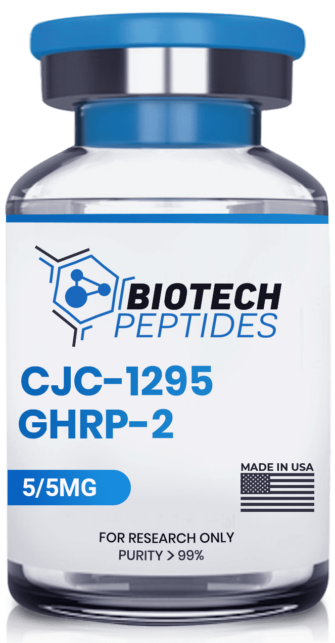
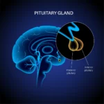
Modified GRF (1-29) is a peptide hormone that is also known as CJC-1295 without DAC (Drug Affinity Complex). It is a synthetic analog of growth hormone-releasing hormone (GHRH) and is designed to increase natural production of growth hormone. Similarly to other GHRH analogs, it appears to work by binding to the growth hormone-releasing hormone receptor in the pituitary gland, and stimulating the release of growth hormone (HGH). Modified GRF (1-29) has been studied for its potential action in research on various conditions, including growth hormone deficiency, osteoporosis, and muscle wasting.
 

Overview of Modified GRF (1-29)

Modified GRF (1-29) is a modified version of GRF (1-29), the smallest amino-acid sequence from the original GHRH that may still retain the ability to trigger the receptors in the pituitary gland and induce an hGH spike. As the name suggests, GRF (1-29) and modified GRF (1-29) are made of 29 amino acids. However, Modified GRF (1-29) has 4 of the original 29 amino acids replaced with the intention to make the peptide more resistant to rapid cleavage by the enzyme dipeptidyl peptidase-4. This cleavage was previously reported to result in peptide inactivation.[1] More specifically, the replaced amino acids are the 2nd, 8th, 15th, and 27th amino acids.

Due to this replacement, the peptide is also known as tetrasubstituted GRF (1-29), and its sequence is Tyr-D-Ala-Asp-Ala-Ile-Phe-Thr-Gln-Ser-Tyr-Arg-Lys-Val-Leu-Ala-Gln-Leu-Ser-Ala-Arg-Lys-Leu-Leu-Gln-Asp-Ile-Leu-Ser-Arg. As a result, the half-life of the peptide has been anticipated to last from 10 – 30 mins.  One of the main modifications is the replacement of L-alanine with D-alanine in the 2nd spot.[2] According to the researchers, “the disappearance half-time of the D-Ala2 analog was 6.7 +/- 0.5, whereas that of GHRH-(1-29)-NH2 was 4.3 +/- 1.4 min (P < 0.05). These findings [suggest] that the D-Ala2 substitution contributes to the enhancement of biological activity by reducing metabolic clearance.


Modified GRF (1-29) and Growth Hormone Synthesis

Modified GRF (1-29) was developed to have a longer half-life but retain identical potential to GRF (1-29), also known as Sermorelin. Since no studies investigate the effects of Modified GRF (1-29) without DAC, the following reports will be based on GRF (1-29) research. Researchers have reported GRF (1-29) to have the potential to increase HGH pulse synthesis.[3] Repeated dosing may lead to a significant increase in growth hormone levels.

A study investigating the action of Modified GRF (1-29) found that it might lead to significant improvements in muscular contractile force.[8] Decline and alterations in muscle mass may result in decreased mobility, and interventions such as GRF (1-29) therapy may potentially mitigate the rapidity or onset of muscular atrophy. Another growth hormone-related impact may be the promotion of skin cell proliferation and increased collagen deposition within the derma.


Modified GRF (1-29) and Composition

One study focused on the impact of Modified GRF (1-29) exposure reported the peptide’s potential to increase growth velocity by 74%, maintained for up to a year of exposure studies.[4] Further studies suggest that exposure to GRF (1-29) for a duration of 4 months resulted in a significant increase in lean body mass. However, it is important to note that such effects were only observed in male research models and were not observed in females.[5] This could be attributed to the fact that GRF (1-29) may not cause growth hormone (hGH) peaks to exceed the physiological limits, which may differ between genders due to biological variations. For example, as the growth hormone remains within its physiological limits in female organisms, it may still significantly be impacted by the negative action of estrogens on IGF-1 production, the main anabolic mediator of hGH.[6] Researchers reported that estrogen appears to impact the HGH-IGF-1 axis “by decreasing liver secretion of insulin-like growth factor-I (IGF-I).[7]

Disclaimer: The products mentioned are not intended for human or animal consumption. Research chemicals are intended solely for laboratory experimentation and/or in-vitro testing.  Bodily introduction of any sort is strictly prohibited by law.  All purchases are limited to licensed researchers and/or qualified professionals. All information shared in this article is for educational purposes only.


References


  1. Scarborough R, Gulyas J, Schally AV, Reeves JJ. Analogs of growth hormone-releasing hormone induce release of growth hormone in the bovine. J Anim Sci. 1988 Jun;66(6):1386-92. doi: 10.2527/jas1988.6661386x. PMID: 3135287.
  2. Soule S, King JA, Millar RP. Incorporation of D-Ala2 in growth hormone-releasing hormone-(1-29)-NH2 increases the half-life and decreases metabolic clearance in normal men. J Clin Endocrinol Metab. 1994 Oct;79(4):1208-11. doi: 10.1210/jcem.79.4.7962295. PMID: 7962295.
  3. Achermann JC, Hindmarsh PC, Robinson IC, Matthews DR, Brook CG. The relative roles of continuous growth hormone-releasing hormone (GHRH(1-29)NH2) and intermittent somatostatin(1-14)(SS) in growth hormone (GH) pulse generation: studies in normal and post cranial irradiated individuals. Clin Endocrinol (Oxf). 1999 Nov;51(5):575-85. doi: 10.1046/j.1365-2265.1999.00839.x. PMID: 10594518.
  4. Thorner M, Rochiccioli P, Colle M, Lanes R, Grunt J, Galazka A, Landy H, Eengrand P, Shah S. Once daily subcutaneous growth hormone-releasing hormone therapy accelerates growth in growth hormone-deficient children during the first year of therapy. Geref International Study Group. J Clin Endocrinol Metab. 1996 Mar;81(3):1189-96. doi: 10.1210/jcem.81.3.8772599. PMID: 8772599.
  5. Khorram O, Laughlin GA, Yen SS. Endocrine and metabolic effects of long-term administration of [Nle27]growth hormone-releasing hormone-(1-29)-NH2 in age-advanced men and women. J Clin Endocrinol Metab. 1997 May;82(5):1472-9. doi: 10.1210/jcem.82.5.3943. PMID: 9141536.
  6. Cook DM. Growth hormone and estrogen: a clinician’s approach. J Pediatr Endocrinol Metab. 2004 Sep;17 Suppl 4:1273-6. PMID: 15506073.
  7. Corpas E, Harman SM, Piñeyro MA, Roberson R, Blackman MR. Growth hormone (GH)-releasing hormone-(1-29) twice daily reverses the decreased GH and insulin-like growth factor-I levels in old men. J Clin Endocrinol Metab. 1992 Aug;75(2):530-5. doi: 10.1210/jcem.75.2.1379256. PMID: 1379256.
  8. Vittone J, Blackman MR, Busby-Whitehead J, Tsiao C, Stewart KJ, Tobin J, Stevens T, Bellantoni MF, Rogers MA, Baumann G, Roth J, Harman SM, Spencer RG. Effects of single nightly injections of growth hormone-releasing hormone (GHRH 1-29) in healthy elderly men. Metabolism. 1997 Jan;46(1):89-96. doi: 10.1016/s0026-0495(97)90174-8. PMID: 9005976.
  9. Khorram O, Laughlin GA, Yen SS. Endocrine and metabolic effects of long-term administration of [Nle27]growth hormone-releasing hormone-(1-29)-NH2 in age-advanced men and women. J Clin Endocrinol Metab. 1997 May;82(5):1472-9. doi: 10.1210/jcem.82.5.3943. PMID: 9141536.
Research Applications of the ARA-290 Peptide

Research Applications of the ARA-290 Peptide

ARA-290 peptide is a small, synthetic peptide is a derivative of erythropoietin (EPO), a hormone that is considered to stimulate the production of red blood cells. However, unlike EPO, ARA-290 does not appear to affect erythropoiesis and has been suggested to exhibit tissue-protective characteristics. In this article, we will explore what ARA-290 peptide is, how it works, and its potential research applications.
 

ARA-290 Peptide Overview

ARA-290 is a synthetic peptide first developed by a research team led by Dr. Schmidt at the New York University School of Medicine. The team discovered the innate repair receptor and suggested that activating it with a synthetic peptide may improve tissue repair and reduce inflammation in various animal disease models.

After further testing and refinement, they developed ARA-290 peptide as a potential agent within the context of research in various conditions involving tissue damage, inflammation, and neuropathic pain. It consists of 11 amino acids designed to mimic a specific sequence of erythropoietin. The amino acid sequence of ARA-290 peptide is Tyr-Glu-Pro-Pro-Pro-Tyr-Gly-Gly-Lys-Pro-Ala

This sequence was identified as the “receptor-specific” site of erythropoietin (EPO), which interacts with a specific receptor on the surface of cells to elicit tissue-protective effects. ARA-290 peptide was developed to specifically target this receptor and avoid interactions with the EPO receptor, which is responsible for the hormone’s potential on erythropoiesis.


ARA-290 Studies on Sarcoidosis-Associated Small Nerve Fiber Loss and Neuropathic Pain

Sarcoidosis-associated small nerve fiber loss and damage (SNFLD) refers to a condition in which the small nerve fibers in the peripheral nervous system are damaged or lost due to sarcoidosis. These are the fibers responsible for transmitting sensory information, such as pain, temperature, and touch. Sarcoidosis is a systemic autoimmune inflammatory disease that may affect multiple organs, including the nerves. It appears to lead to the formation of tiny clusters of inflammatory cells called granulomas, which can cause tissue damage and organ dysfunction.

ARA-290 peptide was hypothesized to reduce symptoms in animal neuropathy models, which led to investigating its potential in research of neuropathy due to Sarcoidosis-associated SNFLD.[1] According to the research, these potential actions may likely be due to the anti-inflammatory characteristics of the peptide that it may potentially exert in nervous tissues. The scientists concluded that “ARA290 … reduced allodynia coupled to suppression of the spinal microglia response, suggestive of a mechanistic link between ARA290-induced suppression of central inflammation and relief of neuropathic pain symptoms.”[2]


ARA-290 Peptide and Diabetes and Related Complications

Insulin resistance plays a significant role in the development and progression of type 2 diabetes. Preliminary research on the glycemic and metabolic potential of ARA-290 peptide was conducted in mice models of insulin resistance, hyperlipidemia, hepatic lipid accumulation, and impaired insulin signaling pathways in skeletal muscle.[5] A high-fat, high-sucrose diet-induced the condition, and exposure to ARA-290 peptide appeared to reduce hepatic lipid deposition and normalized serum glucose and lipid profiles. In this murine model, the exposure appeared to improve insulin sensitivity and glucose uptake in skeletal muscle, attenuate the overproduction of myokines, and enhance mitochondrial biogenesis in skeletal muscle. Many other animal studies also report that ARA-290 peptide may significantly improve glycemic control and lower insulin resistance in rats with type 2 diabetes.[6]

Further studies also report similar results in models of diabetes. According to one study of various diabetes models (mainly type 2 diabetes) and diabetic macular edema (DME), 12 weeks of ARA-290 peptide exposure appeared to lead to improvements in central subfield retinal thickness, tear production, diabetic control, and albuminuria.[7]

Another study included research models of type 2 diabetes who received ARA-290 peptide or a placebo for 28 days and were examined for a month following discontinuation.[8] The researchers suggested that ARA-290 appeared to exhibit an improvement in HbA1c and lipid profiles. They noted that “subjects receiving ARA 290 exhibited an improvement in hemoglobin A1c (Hb A1c) and lipid profiles throughout the 56 [day] observation period.” Neuropathic symptoms and mean corneal nerve fiber density also appeared to improve in the ARA-290 peptide group.


ARA-290 Peptide and Inflammation, Cell Aging

Studies have suggested that ARA-290 peptide may reduce inflammation by modulating the activity of the innate immune system, which is considered responsible for the initial response to infections and tissue damage. Specifically, ARA-290 peptide has been suggested to inhibit the activation of macrophages and reduce the production of pro-inflammatory cytokines, such as TNF-alpha and IL-1beta, in models of colon inflammation.[9]

One trial in aged rats reports that chronic ARA-290 peptide exposure may possibly reduce inflammation and fibrosis in the heart, improve mitochondrial and myocardial cell function, and preserve left ventricular ejection fraction.[10] ARA-290 also apparently mitigated the age-associated increase in blood pressure, preserved body weight, and reduced markers of organism-wide frailty.


Conclusion

More research is needed to fully understand ARA-290’s potential.  Currently, the peptide has research-only status, and it is not intended for  managing any condition.

Disclaimer: The products mentioned are not intended for human or animal consumption. Research chemicals are intended solely for laboratory experimentation and/or in-vitro testing.  Bodily introduction of any sort is strictly prohibited by law.  All purchases are limited to licensed researchers and/or qualified professionals. All information shared in this article is for educational purposes only.


References


  1. Swartjes M, van Velzen M, Niesters M, Aarts L, Brines M, Dunne A, Cerami A, Dahan A. ARA 290, a peptide derived from the tertiary structure of erythropoietin, produces long-term relief of neuropathic pain coupled with suppression of the spinal microglia response. Mol Pain. 2014 Feb 16;10:13. doi: 10.1186/1744-8069-10-13. PMID: 24529189; PMCID: PMC3928087.
  2. Dahan A, Dunne A, Swartjes M, Proto PL, Heij L, Vogels O, van Velzen M, Sarton E, Niesters M, Tannemaat MR, Cerami A, Brines M. ARA 290 improves symptoms in patients with sarcoidosis-associated small nerve fiber loss and increases corneal nerve fiber density. Mol Med. 2013 Nov 8;19(1):334-45. doi: 10.2119/molmed.2013.00122. Erratum in: Mol Med. 2016 Oct 20;22:674. PMID: 24136731; PMCID: PMC3883966.
  3. Culver DA, Dahan A, Bajorunas D, Jeziorska M, van Velzen M, Aarts LPHJ, Tavee J, Tannemaat MR, Dunne AN, Kirk RI, Petropoulos IN, Cerami A, Malik RA, Brines M. Cibinetide Improves Corneal Nerve Fiber Abundance in Patients With Sarcoidosis-Associated Small Nerve Fiber Loss and Neuropathic Pain. Invest Ophthalmol Vis Sci. 2017 May 1;58(6):BIO52-BIO60. doi: 10.1167/iovs.16-21291. PMID: 28475703.
  4. Heij L, Niesters M, Swartjes M, Hoitsma E, Drent M, Dunne A, Grutters JC, Vogels O, Brines M, Cerami A, Dahan A. Safety and efficacy of ARA 290 in sarcoidosis patients with symptoms of small fiber neuropathy: a randomized, double-blind pilot study. Mol Med. 2012 Nov 15;18(1):1430-6. doi: 10.2119/molmed.2012.00332. PMID: 23168581; PMCID: PMC3563705.
  5. Collino M, Benetti E, Rogazzo M, Chiazza F, Mastrocola R, Nigro D, Cutrin JC, Aragno M, Fantozzi R, Minetto MA, Thiemermann C. A non-erythropoietic peptide derivative of erythropoietin decreases susceptibility to diet-induced insulin resistance in mice. Br J Pharmacol. 2014 Dec;171(24):5802-15. doi: 10.1111/bph.12888. Epub 2014 Nov 24. PMID: 25164531; PMCID: PMC4290718.
  6. Muller C, Yassin K, Li LS, Palmblad M, Efendic S, Berggren PO, Cerami A, Brines M, Östenson CG. ARA290 Improves Insulin Release and Glucose Tolerance in Type 2 Diabetic Goto-Kakizaki Rats. Mol Med. 2016 May;21(1):969-978. doi: 10.2119/molmed.2015.00267. Epub 2015 Dec 29. PMID: 26736179; PMCID: PMC4818260.
  7. Lois N, Gardner E, McFarland M, Armstrong D, McNally C, Lavery NJ, Campbell C, Kirk RI, Bajorunas D, Dunne A, Cerami A, Brines M. A Phase 2 Clinical Trial on the Use of Cibinetide for the Treatment of Diabetic Macular Edema. J Clin Med. 2020 Jul 14;9(7):2225. doi: 10.3390/jcm9072225. PMID: 32674280; PMCID: PMC7408632.
  8. Brines M, Dunne AN, van Velzen M, Proto PL, Ostenson CG, Kirk RI, Petropoulos IN, Javed S, Malik RA, Cerami A, Dahan A. ARA 290, a nonerythropoietic peptide engineered from erythropoietin, improves metabolic control and neuropathic symptoms in patients with type 2 diabetes. Mol Med. 2015 Mar 13;20(1):658-66. doi: 10.2119/molmed.2014.00215. PMID: 25387363; PMCID: PMC4365069.
  9. Nairz M, Haschka D, Dichtl S, Sonnweber T, Schroll A, Aßhoff M, Mindur JE, Moser PL, Wolf D, Swirski FK, Theurl I, Cerami A, Brines M, Weiss G. Cibinetide dampens innate immune cell functions thus ameliorating the course of experimental colitis. Sci Rep. 2017 Oct 12;7(1):13012. doi: 10.1038/s41598-017-13046-3. PMID: 29026145; PMCID: PMC5638901.
  10. Winicki NM, Nanavati AP, Morrell CH, Moen JM, Axsom JE, Krawczyk M, Petrashevskaya NN, Beyman MG, Ramirez C, Alfaras I, Mitchell SJ, Juhaszova M, Riordon DR, Wang M, Zhang J, Cerami A, Brines M, Sollott SJ, de Cabo R, Lakatta EG. A small erythropoietin derived non-hematopoietic peptide reduces cardiac inflammation, attenuates age associated declines in heart function and prolongs healthspan. Front Cardiovasc Med. 2023 Jan 18;9:1096887. doi: 10.3389/fcvm.2022.1096887. PMID: 36741836; PMCID: PMC9889362.
Sermorelin Peptide Potential as a Growth Hormone Secretagogue

Sermorelin Peptide Potential as a Growth Hormone Secretagogue

Sermorelin Acetate, a synthetic peptide, was developed to function as an analog of a naturally occurring hormone called growth hormone-releasing hormone (GHRH). While GHRH has 44 amino acids, Sermorelin peptide is truncated down to 29 amino acids. Sermorelin peptide is also known as the Growth Hormone Releasing Factor (1-29) or GRF (1-29). Sermorelin is the smallest fragment of the GHRH chain that appears to possess the same functions, namely, stimulating the pulsatile release of growth hormone (HGH) by the pituitary gland. Due to this potential, Sermorelin peptide has been classified by researchers as a Growth Hormone Secretagogue (GHS). Like GHRH and other GHSs, Sermorelin may possibly increase the production of HGH in the pituitary gland, resulting in higher and more frequent peaks of the growth hormones. This would also result in elevated levels of Insulin Growth Factor-1 (IGF-1), a primary anabolic mediator.
 

What is the Sermorelin?

Sermorelin was first synthesized in the early 1980s and has been avidly researched since its development. It is made of 29 amino acids and bears the sequence YADAXFXNSYRKVLGQLSARKLLQDXMSR. Sermorelin peptide, aka GRF (1-29), should not be confused with Modified GRF (1-29), which has 4 of the original 29 amino acids replaced with the intent to increase its half-life from 10 to 30 minutes.


General Research in Sermorelin


Sermorelin Peptide and the Endocrine System

As a GHRH analog, Sermorelin peptide has been researched primarily for its potential to stimulate pulsatile hGH synthesis via the pituitary gland. Studies observed that as long as the pituitary gland is functioning correctly, Sermorelin, combined with the amino acid Arginine, may induce a significant spike in serum HGH levels.[1] Studies in research models of growth failure also report that Sermorelin peptide appeared to increase growth velocity with growth failure and functional pituitary glands by 74%.[2] Further research reported that 6 months of either Sermorelin peptide or hGH exposure, appeared to exhibit a similar increase in growth velocity.[3] Studies also reported an apparent significant increase in serum HGH levels after several days of Sermorelin peptide introduction.[4]

The peptide may also affect the levels of other hormones apart from HGH, such as insulin and sex hormones.  Although high hGH is often associated with increased insulin levels and insulin resistance, one 16-week trial suggested that Sermorelin may improve insulin sensitivity in males but not in female organisms.[5]  One animal trial reported an increase in gonadotropic hormones and testosterone levels in male mice, but there is no clinical research to support these findings.[6]


Sermorelin Studies in Cardiovascular Function

Sermorelin may impact cardiovascular functioning due to its apparent HGH-stimulating potential.[7] Researchers suggest, “That GHRH analog [exposure] induced anabolic effects favoring [male species] more than [female]. Further studies are needed to define the gender differences observed in response to GHRH analog [exposure].[8]


Sermorelin Peptide and Extracellular Matrix

Scientists posit that Sermorelin has the potential to function in increasing HGH levels. From this assumption, it may be possible for Sermorelin peptide to increase the proliferation of new skin cells, ultimately leading to increased collagen deposition inside the derma.

One study focusing on male species reported that 4 months of Sermorelin peptide exposure appeared to significantly increase their skin thickness.[11] Additionally, the males, but not the female organisms, also expressed increased libido and related behaviors.


Sermorelin Peptide and the Nervous System

One study suggested that even a one-time exposure to Sermorelin peptide may support improvements in short-term memory.[12] In fact, researchers reported that the effect on memory recall appeared more significant in Sermorelin-exposed research models than those given a placebo. There is also mixed data regarding the potential of Sermorelin peptide on brain tumors. One experiment reports that Sermorelin peptide may speed up the development of neuroendocrine tumors such as pituitary adenomas due to its possible growth-promoting effect.[13] However, a study in gliomas reported the opposite effect, observing that the peptide appeared to suppress the recurrence of the tumors.[14]


Conclusion

Sermorelin may hold significant research potential as a Growth Hormone Secretagogue (GSH). This is supported by research findings indicating that when exposed to Sermorelin, animal research models’ serum growth hormone levels might not exceed the physiological limits exerted by somatostatin.

Disclaimer: The products mentioned are not intended for human or animal consumption. Research chemicals are intended solely for laboratory experimentation and/or in-vitro testing.  Bodily introduction of any sort is strictly prohibited by law.  All purchases are limited to licensed researchers and/or qualified professionals. All information shared in this article is for educational purposes only.


References


  1. Prakash A, Goa KL. Sermorelin: a review of its use in the diagnosis and treatment of children with idiopathic growth hormone deficiency. BioDrugs. 1999 Aug;12(2):139-57. doi: 10.2165/00063030-199912020-00007. PMID: 18031173.
  2. Thorner M, Rochiccioli P, Colle M, Lanes R, Grunt J, Galazka A, Landy H, Eengrand P, Shah S. Once daily subcutaneous growth hormone-releasing hormone therapy accelerates growth in growth hormone-deficient children during the first year of therapy. Geref International Study Group. J Clin Endocrinol Metab. 1996 Mar;81(3):1189-96. doi: 10.1210/jcem.81.3.8772599. PMID: 8772599.
  3. Neyzi O, Yordam N, Ocal G, Bundak R, Darendeliler F, Açikgöz E, Berberoğlu M, Günöz H, Saka N, Calikoğlu AS. Growth response to growth hormone-releasing hormone(1-29)-NH2 compared with growth hormone. Acta Paediatr Suppl. 1993 Mar;388:16-21; discussion 22. doi: 10.1111/j.1651-2227.1993.tb12828.x. PMID: 8329826.
  4. Achermann JC, Hindmarsh PC, Robinson IC, Matthews DR, Brook CG. The relative roles of continuous growth hormone-releasing hormone (GHRH(1-29)NH2) and intermittent somatostatin(1-14)(SS) in growth hormone (GH) pulse generation: studies in normal and post cranial irradiated individuals. Clin Endocrinol (Oxf). 1999 Nov;51(5):575-85. doi: 10.1046/j.1365-2265.1999.00839.x. PMID: 10594518.
  5. Khorram O, Laughlin GA, Yen SS. Endocrine and metabolic effects of long-term administration of [Nle27]growth hormone-releasing hormone-(1-29)-NH2 in age-advanced men and women. J Clin Endocrinol Metab. 1997 May;82(5):1472-9. doi: 10.1210/jcem.82.5.3943. PMID: 9141536.
  6. Chatelain PG, Sanchez P, Saez JM. Growth hormone and insulin-like growth factor I treatment increase testicular luteinizing hormone receptors and steroidogenic responsiveness of growth hormone deficient dwarf mice. Endocrinology. 1991 Apr;128(4):1857-62. doi: 10.1210/endo-128-4-1857. PMID: 2004605.
  7. Khorram O, Laughlin GA, Yen SS. Endocrine and metabolic effects of long-term administration of [Nle27]growth hormone-releasing hormone-(1-29)-NH2 in age-advanced men and women. J Clin Endocrinol Metab. 1997 May;82(5):1472-9. doi: 10.1210/jcem.82.5.3943. PMID: 9141536.
  8. Vittone J, Blackman MR, Busby-Whitehead J, Tsiao C, Stewart KJ, Tobin J, Stevens T, Bellantoni MF, Rogers MA, Baumann G, Roth J, Harman SM, Spencer RG. Effects of single nightly injections of growth hormone-releasing hormone (GHRH 1-29) in healthy elderly men. Metabolism. 1997 Jan;46(1):89-96. doi: 10.1016/s0026-0495(97)90174-8. PMID: 9005976.
  9. Corpas E, Harman SM, Piñeyro MA, Roberson R, Blackman MR. Growth hormone (GH)-releasing hormone-(1-29) twice daily reverses the decreased GH and insulin-like growth factor-I levels in old men. J Clin Endocrinol Metab. 1992 Aug;75(2):530-5. doi: 10.1210/jcem.75.2.1379256. PMID: 1379256.
  10. Vittone J, Blackman MR, Busby-Whitehead J, Tsiao C, Stewart KJ, Tobin J, Stevens T, Bellantoni MF, Rogers MA, Baumann G, Roth J, Harman SM, Spencer RG. Effects of single nightly injections of growth hormone-releasing hormone (GHRH 1-29) in healthy elderly men. Metabolism. 1997 Jan;46(1):89-96. doi: 10.1016/s0026-0495(97)90174-8. PMID: 9005976.
  11. Khorram O, Laughlin GA, Yen SS. Endocrine and metabolic effects of long-term administration of [Nle27]growth hormone-releasing hormone-(1-29)-NH2 in age-advanced men and women. J Clin Endocrinol Metab. 1997 May;82(5):1472-9. doi: 10.1210/jcem.82.5.3943. PMID: 9141536.
  12. Alvarez XA, Cacabelos R. Effects of GRF (1-29) NH2 on short-term memory: neuroendocrine and neuropsychological assessment in healthy young subjects. Methods Find Exp Clin Pharmacol. 1990 Sep;12(7):493-9. PMID: 2087150.
  13. Stepień T, Sacewicz M, Lawnicka H, Krupiński R, Komorowski J, Siejka A, Stepień H. Stimulatory effect of growth hormone-releasing hormone (GHRH(1-29)NH2) on the proliferation, VEGF and chromogranin A secretion by human neuroendocrine tumor cell line NCI-H727 in vitro. Neuropeptides. 2009 Oct;43(5):397-400. doi: 10.1016/j.npep.2009.08.005. Epub 2009 Sep 10. PMID: 19747727.
  14. Chang Y, Huang R, Zhai Y, Huang L, Feng Y, Wang D, Chai R, Zhang W, Hu H. A potentially effective drug for patients with recurrent glioma: sermorelin. Ann Transl Med. 2021 Mar;9(5):406. doi: 10.21037/atm-20-6561. PMID: 33842627; PMCID: PMC8033379.