Vialox Peptide: Neuromuscular Transmission and Skin Wrinkle Reduction
By potentially diminishing the release of acetylcholine, Vialox may contribute to muscle relaxation, thereby possibly reducing the depth and formation of wrinkles along the skin barrier. The proposed mechanism of action for Vialox bears resemblance to that of tubocurarine, a natural alkaloid compound known for its muscle-relaxing properties.
Featured Product
 (200mg)")
Scientific and Research Studies
Action Mechanisms
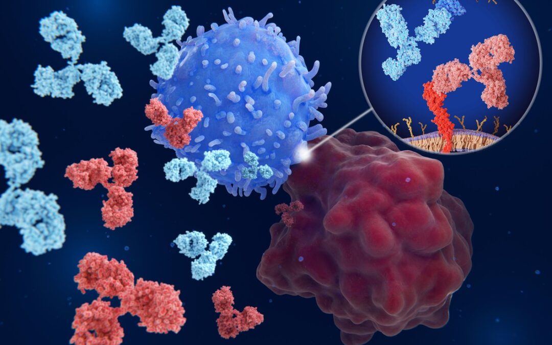
Vialox is suggested by researchers to inhibit neurotransmitter activity.[2] It may function similarly to tubocurarine through its interaction with acetylcholine receptors on the postsynaptic membrane of muscle cells.[3] Tubocurarine, a naturally occurring alkaloid found in the bark of plants such as Chondrodendron tomentosum (commonly known as “curare”), is considered a potent neurotoxin. It acts as a non-depolarizing neuromuscular blocker by preventing acetylcholine from inducing muscle contraction at the neuromuscular junction.
Researchers also classify Vialox as a non-depolarizing neuromuscular blocker. The peptide binds to acetylcholine receptors on the postsynaptic membrane of muscle cells, acting as “a competitive antagonist” at these receptor sites.[4] It may interact with neuronal nicotinic acetylcholine receptors, which play a role in regulating muscle contraction by mediating communication between motor nerves and muscles at the neuromuscular junction.
As an antagonist, Vialox appears to block acetylcholine binding to these receptors, preventing the opening of sodium ion channels required for depolarizing the cell and initiating muscle contraction.[5] By inhibiting acetylcholine receptor activity, Vialox may potentially keep smooth muscles relaxed, possibly reducing wrinkles in the skin barrier as a result.
Vialox Peptide and Wrinkle Formation, Skin Texture
Research into Vialox has explored its potential to mitigate wrinkles on the skin surface and decrease texture variations along the skin barrier. The study of compounds in wrinkle reduction carries certain risks, particularly with higher concentrations or prolonged exposure. Long-term exposure may also pose unknown or unanticipated ancillary impacts within the laboratory test models. However, Vialox is noted for its short half-life and potential for less abrasive impacts. Studies suggest that this compound may cause “softened wrinkles and reduced skin roughness.”[5]
Experimental data indicated a reduction in muscle contractions by 71% within one minute of Vialox exposure, with a subsequent 58% reduction observed after two hours. These findings imply that the decreased frequency of muscle contractions may contribute to the formation of shallower lines on the skin’s surface.
Additional research supports Vialox’s potential in reducing the development and depth of skin wrinkles.[7] Results indicate a 49% decrease in wrinkle size and a 47% reduction in skin roughness after 28 days of consistent exposure.
Vialox Peptide and Neuromuscular Transmission
Vialox has garnered scientific interest due to its proposed ability to disrupt nerve-muscle communication. Unlike other antagonists of nicotinic acetylcholine receptors (AChR), Vialox appears to act exclusively on peripheral AChR, with minimal impact on central neuronal receptors as suggested by animal studies. Researchers hypothesize that Vialox might be effective in addressing certain spastic conditions, including migraines and muscle spasms.
Vialox is suggested to interfere with signal transmission between neurons and muscles by acting as an antagonist of the acetylcholine receptor. It appears to block nerve signals at the post-synaptic membrane, leading to muscle relaxation. Normally, when a nerve’s axon releases acetylcholine, these signals travel to the neuromuscular junction and bind to receptors on the muscle, facilitating the release of sodium ions. This process may result in depolarization, generating the electrical pulse responsible for muscle contraction and wrinkle formation.
Vialox is studied for its potential to halt this process by binding to AChR, thereby preventing acetylcholine from attaching to these receptors. This blockade is thought to reduce both the frequency and intensity of muscular contractions, similar to the effects induced by botulinum toxin, tubocurarine, and curare toxin. The consequent partial paralysis of muscles leads to forced relaxation.
Disclaimer: The products mentioned are not intended for human or animal consumption. Research chemicals are intended solely for laboratory experimentation and/or in-vitro testing. Bodily introduction of any sort is strictly prohibited by law. All purchases are limited to licensed researchers and/or qualified professionals. All information shared in this article is for educational purposes only.
References:
- National Center for Biotechnology Information (2024). PubChem Compound Summary for CID 67073230, Vialox. Retrieved August 4, 2024 from https://pubchem.ncbi.nlm.nih.gov/compound/Vialox.
- Husein el Hadmed, H., & Castillo, R. F. (2016). Cosmeceuticals: peptides, proteins, and growth factors. Journal of cosmetic dermatology, 15(4), 514-519. Husein el Hadmed, H., & Castillo, R. F. (2016). Cosmeceuticals: peptides, proteins, and growth factors. Journal of cosmetic dermatology, 15(4), 514-519. https://pubmed.ncbi.nlm.nih.gov/27142709/
- Lupo, M. P., & Cole, A. L. (2007). Cosmeceutical peptides. Dermatologic therapy, 20(5), 343-349. https://pubmed.ncbi.nlm.nih.gov/18045359/
- Gorouhi, F., & Maibach, H. I. (2009). Role of peptides in preventing or treating aged skin. International journal of cosmetic science, 31(5), 327-345. https://pubmed.ncbi.nlm.nih.gov/19570099/
- Satriyasa B. K. (2019). Botulinum toxin (Botox) A for reducing the appearance of facial wrinkles: a literature review of clinical use and pharmacological aspect. Clinical, cosmetic and investigational dermatology, 12, 223–228. https://doi.org/10.2147/CCID.S202919
- Kalandakanond, S., & Coffield, J. A. (2001). Cleavage of SNAP-25 by botulinum toxin type A requires receptor-mediated endocytosis, pH-dependent translocation, and zinc. The Journal of pharmacology and experimental therapeutics, 296(3), 980–986. https://pubmed.ncbi.nlm.nih.gov/11181932/
- Reddy BY, Jow T, Hantash BM. Bioactive oligopeptides in dermatology: Part II. Exp Dermatol. 2012 Aug;21(8):569-75. doi: 10.1111/j.1600-0625.2012.01527.x. Epub 2012 Jun 4. PMID: 22672721. https://pubmed.ncbi.nlm.nih.gov/22672721/
- Lebedev DS, Kryukova EV, Ivanov IA, Egorova NS, Timofeev ND, Spirova EN, Tufanova EY, Siniavin AE, Kudryavtsev DS, Kasheverov IE, Zouridakis M, Katsarava R, Zavradashvili N, Iagorshvili I, Tzartos SJ, Tsetlin VI. Oligoarginine Peptides, a New Family of Nicotinic Acetylcholine Receptor Inhibitors. Mol Pharmacol. 2019 Nov;96(5):664-673. doi: 10.1124/mol.119.117713. Epub 2019 Sep 6. PMID: 31492697. https://pubmed.ncbi.nlm.nih.gov/31492697/
")

")
")


")