
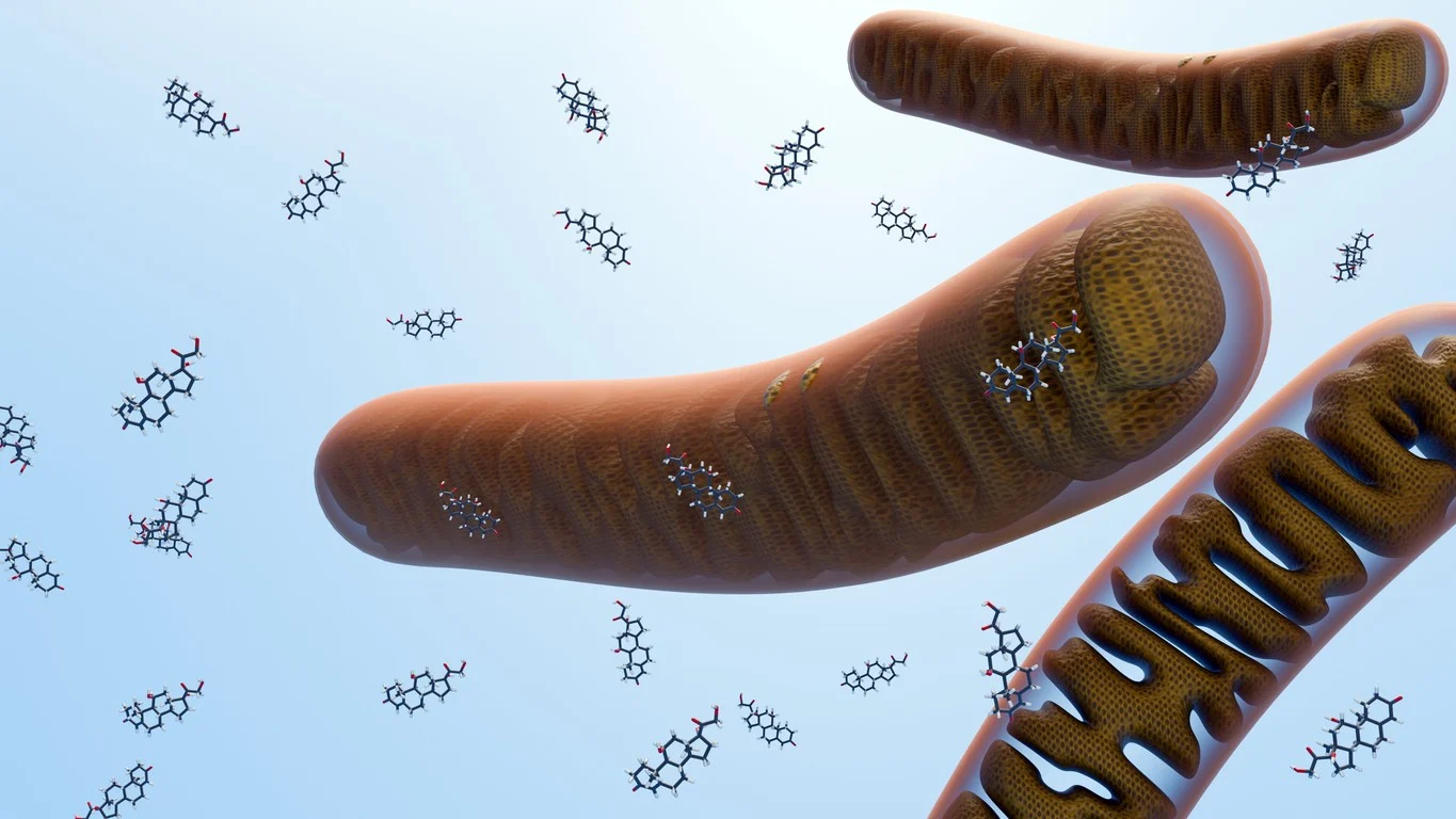
GHK-Cu modulates extracellular matrix (ECM) remodeling via matrix metalloproteinase regulation and collagen synthesis.[7][8] KPV mitigates NF-κB-dependent inflammatory signaling through intracellular uptake mediated by the PepT1 transporter.[10] This convergent but non-redundant receptor architecture may provide a research framework for studying multi-pathway tissue biology, inflammation modulation, and cellular regeneration across diverse preclinical models.
Contents:
- KLOW Blend Historical Development
- KLOW Blend Proposed Mechanism of Action
- KLOW Blend Scientific and Research Studies
- BPC-157: FAK-Paxillin Pathway Activation and Tendon Fibroblast Biology
- BPC-157: VEGFR2-Mediated Angiogenesis and Vascular Signaling
- TB-500 (Thymosin Beta-4): ILK-Akt Activation and Cardiac Cell Survival
- B-500 (Thymosin Beta-4): Epicardial Progenitor Mobilization and Neovascularisation
- GHK-Cu: ECM Remodeling, Collagen Synthesis, and Glycosaminoglycan Production
- GHK-Cu: Anti-Inflammatory Activity in Acute Tissue Injury Models
- KPV: PepT1-Mediated Uptake and NF-κB Mitigation in Intestinal Inflammation Models
- KPV: Melanocortin-Derived Anti-Inflammatory Activity in IBD Models
- References
Featured Product

KLOW Blend Historical Development of Constituent Peptides
BPC-157 was first isolated and characterized from gastric juice by Professor Predrag Sikirić and colleagues at the University of Zagreb, Croatia, in the early 1990s.[1] The compound (CAS 137525-51-0; also designated PL 14736) was identified as a 15-amino acid partial sequence of the endogenous protection compound protein, exhibiting cytoprotective properties in gastric mucosal models. Its stability relative to other bioactive peptides-attributable to its proline-rich central motif-facilitated investigation across a range of murine tissue systems.[3]
TB-500 is a synthetic heptapeptide corresponding to residues 17–23 of full-length thymosin beta-4 (β4), a ubiquitous 43-amino acid G-actin sequestering protein originally isolated from bovine thymus in the 1960s.[4][6] The designation TB-500 refers to the acetylated active fragment Ac-LKKTETQ, which retains the actin-binding motif of the parent molecule. The full-length β4 molecule underpins the preponderance of the published academic literature, with TB-500 used in research contexts to access this actin-regulatory biology.[4]
GHK-Cu was isolated from plasma by Pickart in 1973, originally identified as a factor within the albumin fraction that induced aged liver cells to synthesize proteins with a profile resembling that of younger tissue.[7] Subsequent investigations established that this activity resided in the tripeptide glycyl-L-histidyl-L-lysine, which forms a stable chelate with copper(II) ions (GHK-Cu). Research has since characterized GHK-Cu as a regulator of ECM synthesis, collagen production, and tissue remodeling processes.[7][8]
KPV (Lys-Pro-Val) corresponds to C-terminal residues 11–13 of α-melanocyte-stimulating hormone (α-MSH), a 13-amino acid neuropeptide of pituitary origin. Research investigations identified that this tripeptide fragment retains the anti-inflammatory activity of the parent hormone while lacking its pigmentation-inducing and hunger hormone-modulating implications.[10][11] The small molecular size of KPV may confer oral stability and facilitate intracellular entry via di/tripeptide transporters, properties that have been examined in gastrointestinal and inflammatory research models.
KLOW Blend Receptor Mechanisms and Intracellular Signaling
BPC-157 engages VEGFR2 (vascular endothelial growth factor receptor 2), a receptor tyrosine kinase expressed on endothelial cells. VEGFR2 activation may initiate downstream phosphorylation cascades through the Akt-eNOS axis, promoting nitric oxide (NO) synthesis and endothelial cell proliferation.[2] Separately, BPC-157 has been observed to activate the focal adhesion kinase (FAK)-paxillin pathway in tendon fibroblasts, a signaling axis associated with increased cell migration and cytoskeletal reorganization.[1] Research suggests BPC-157 may also upregulate growth hormone receptor expression in fibroblast populations, potentially potentiating GH-mediated repair signaling.[3]
TB-500’s primary mechanism involves sequestration of monomeric G-actin, maintaining the intracellular pool of actin in a polymerization-competent state and enabling rapid cytoskeletal reorganization during cell migration.[6] Integrin-linked kinase (ILK) activation represents a downstream consequence of TB-500/β4 exposure in cardiac and endothelial cells, initiating Akt-mediated survival signaling.⁵ In cardiac injury models, ILK and Akt upregulation following β4 exposure has been associated with supported early cardiomyocyte survival and neovascularisation.
GHK-Cu engages fibroblasts and dermal cells to upregulate matrix metalloproteinase-2 (MMP-2), an enzyme involved in ECM remodeling and turnover of fibrillar collagen.[8] Concurrently, GHK-Cu may stimulate synthesis of type I collagen, elastin, and sulfated glycosaminoglycans – key structural ECM components. Research suggests these dual MMP-upregulatory and collagen-stimulatory implications might indicate a coordinated ECM remodeling capacity, consistent with its historical characterization as a wound recovery activator.
KPV enters intestinal epithelial cells and immune cells primarily through the PepT1 di/tripeptide transporter, an uptake mechanism that is upregulated in inflamed intestinal tissue during inflammatory bowel conditions. Once internalized, KPV mitigates NF-κB nuclear translocation by attenuating IkB-α degradation, thereby reducing transcriptional activation of pro-inflammatory cytokine genes including IL-8, TNF-α, and IL-1β.[10][11] Research suggests this intracellular NF-κB mitigation may operate independently of melanocortin receptor (MCR) engagement, distinguishing its mechanism from the parent α-MSH molecule.
KLOW Blend Scientific and Research Studies
BPC-157: FAK-Paxillin Pathway Activation and Tendon Fibroblast Biology
Researchers studied the mechanistic basis of BPC-157’s implications on tendon recovery using cultured murine model Achilles tendon fibroblasts and tendon explant outgrowth assays.[1] BPC-157 significantly accelerated the outgrowth of tendon fibroblasts from explant cultures and markedly increased in vitro fibroblast migration in a concentration-dependent manner. Cell proliferation, as measured by MTT assay, was not directly altered, suggesting that the primary implication may involve cell motility and tissue invasion rather than proliferative expansion.
Western blot analysis indicated that BPC-157 produced concentration-dependent increases in the phosphorylation of FAK and paxillin-key components of focal adhesion complexes-without altering total protein levels. FITC-phalloidin staining further suggested supported F-actin formation in BPC-157-treated fibroblasts. Research suggests these findings might indicate that BPC-157 promotes tendon cell migration and structural reorganization through activation of the FAK-paxillin focal adhesion pathway, potentially contributing to observed accelerations in tendon wound recovery in preclinical models.
BPC-157: VEGFR2-Mediated Angiogenesis and Vascular Signaling
Hsieh et al. (2017)[2] examined the angiogenic signaling mechanisms of BPC-157 in endothelial cell models and correlated receptor-level activity with vascular outcomes. Findings suggested that BPC-157 may upregulate VEGFR2 expression in endothelial cells and activate downstream Akt-eNOS phosphorylation cascades, which are associated with endothelial nitric oxide production and angiogenic sprouting. These receptor-level observations were correlated with supported vessel formation markers in the murine models studied.
Brcic et al. (2009)[3] examined the angiogenic potential of BPC-157 across muscle tissue and murine tendon recovery research models, correlating peptide exposure with VEGF expression using cell culture and animal systems. Observations suggested that BPC-157 may stimulate angiogenesis by upregulating VEGF expression at wound sites, with the angiogenic response appearing closely associated with the recovery progression documented histologically across tissue compartments. Research suggests these findings might indicate that VEGFR2-driven angiogenesis represents a mechanistically central element of BPC-157’s tissue repair activity in preclinical settings.
TB-500 (Thymosin Beta-4): ILK-Akt Activation and Cardiac Cell Survival
Bock-Marquette et al. (2004)[5] studied the role of thymosin beta-4 in cardiac cell biology following myocardial injury. In a murine coronary artery ligation model, β4 exposure was associated with upregulation of integrin-linked kinase (ILK) and Akt activity within the cardiac tissue. supported early cardiomyocyte survival was observed in treated animals, alongside support for cardiac functional parameters relative to control groups.
The investigators further characterized β4’s implications on cardiac progenitor cell migration in vitro, finding that the peptide promoted cardiomyocyte migration through ILK-dependent pathways. Research suggests these findings might indicate that β4-mediated ILK-Akt signaling may represent a cardioprotective mechanism with potential relevance to preclinical models of ischemic cardiac injury. These observations provide mechanistic context for TB-500’s G-actin-based biology in cardiac tissue, where cytoskeletal regulation is central to cell migration and structural repair.
TB-500 (Thymosin Beta-4): Epicardial Progenitor Mobilization and Neovascularisation
Smart et al. (2007)[12] examined the capacity of thymosin beta-4 to mobilize adult epicardial progenitor cells and stimulate neovascularisation in a murine cardiac injury model. Following myocardial infarction, β4 exposure was associated with reactivation of dormant epicardial progenitor populations and promotion of new coronary vessel formation. The investigators proposed that β4 may stimulate the re-expression of embryonic epicardial genes, enabling adult epicardial cells to contribute to vascular repair processes.
Actin regulation by the β4 molecule was identified as integral to the progenitor mobilization response, linking the peptide’s primary biochemical function (G-actin sequestration) to downstream biological outcomes in a clinically relevant injury model. Research suggests these findings might indicate that TB-500’s actin-regulatory mechanism may drive paracrine and regenerative signalling beyond the immediate site of peptide engagement.
GHK-Cu: ECM Remodeling, Collagen Synthesis, and Glycosaminoglycan Production
Researchers conducted a comprehensive review[7] of GHK-Cu’s biological activity across dermatology and wound biology models. Investigations cited therein indicated that GHK-Cu may stimulate collagen synthesis in fibroblast cultures, accelerate wound contraction and epithelialization, and promote the take of transplanted dermal cells in murine models. The review further identified GHK-Cu’s capacity to upregulate MMP-2 expression, facilitating ECM remodeling and clearance of damaged matrix proteins. Endogenous plasma GHK concentrations of approximately 200 ng/mL at age 20 were reported to decline to approximately 80 ng/mL by age 60, with this decline noted to correlate with observed reductions in tissue regenerative capacity.
Scientists[8] examined the role of matrikines-ECM-derived peptide fragments including GHK-in the regulation of ECM degradation and synthesis. Findings supported the characterization of GHK as a matrikine capable of modulating MMP expression and promoting glycosaminoglycan synthesis in wound tissue models, implications potentially relevant to the maintenance of structural ECM integrity during repair processes. Research suggests these findings might indicate that GHK-Cu may function as an endogenous ECM regulatory signal with broad relevance to tissue homeostasis research.
GHK-Cu: Anti-Inflammatory Activity in Acute Tissue Injury Models
Research was conducted studying the implications of GHK-Cu in a murine model of lipopolysaccharide (LPS)-induced acute lung injury (ALI). GHK-Cu exposure was associated with suppression of pro-inflammatory mediators including reactive oxygen species (ROS), nitric oxide (NO), and cyclooxygenase-2 (COX-2) in pulmonary tissue. Reductions in transforming growth factor-beta (TGF-β) and tumor necrosis factor-alpha (TNF-α) concentrations were also reported in treated animals relative to untreated controls.[9]
Histopathological evaluation of lung tissue suggested reduced inflammatory infiltrate and structural preservation in GHK-Cu-exposed groups. Research suggests these observations might indicate that GHK-Cu may engage anti-inflammatory pathways beyond its ECM-regulatory function, potentially through modulation of iron-mediated oxidative stress and cytokine regulatory networks. These findings may complement the anti-inflammatory mechanism of KPV within the KLOW blend by providing an additional, mechanistically distinct pathway of inflammatory modulation.
KPV: PepT1-Mediated Uptake and NF-κB Mitigation in Intestinal Inflammation Models
Dalmasso et al. (2008)[10] studied the mechanism underlying KPV’s anti-inflammatory activity in intestinal epithelial and immune cell models. Using intestinal epithelial cell lines (Caco2-BBE and HT29-Cl.19A) and Jurkat T cells stimulated with pro-inflammatory cytokines, the investigators examined whether KPV’s anti-inflammatory implication required the PepT1 di/tripeptide transporter. Uptake experiments using radiolabelled [3H]KPV and competitive mitigation assays confirmed PepT1-mediated intracellular entry in PepT1-expressing cells.
Mechanistic assays indicated that KPV attenuated NF-κB-dependent gene expression by mitigating IkB-α degradation, with downstream reductions in IL-8 mRNA expression. In two murine colitis models (DSS-induced and CD45RBhi transfer colitis), systemic KPV exposure was associated with attenuation of colonic inflammation, as assessed by histological scoring and cytokine measurement. Research suggests these findings might indicate that PepT1-mediated intracellular delivery may be a prerequisite for KPV’s anti-inflammatory activity in epithelial tissues, and that upregulation of PepT1 during intestinal inflammation may support local peptide uptake at inflamed mucosal sites.
KPV: Melanocortin-Derived Anti-Inflammatory Activity in IBD Models
Scientists evaluated KPV’s potential in two established murine intestinal inflammation models: DSS-induced colitis and CD45RBhi transfer colitis. The study additionally examined KPV activity in animals carrying a non-functional melanocortin-1 receptor (MC1Re/e), enabling dissection of receptor-dependent from receptor-independent anti-inflammatory mechanisms.[11]
Findings suggested that KPV attenuated clinical and histological indices of colitis in both models, and that this anti-inflammatory activity was preserved in MC1R-deficient animals. Research suggests these results might indicate that KPV’s gastrointestinal anti-inflammatory mechanism may not require functional MCR engagement, consistent with the NF-κB-centric, receptor-independent intracellular pathway characterized by Dalmasso et al.[11] The mechanistic independence of KPV from its parent hormone’s receptor may have implications for the design of research models investigating targeted mucosal anti-inflammatory signaling.
Disclaimer: The products mentioned are not intended for human or animal consumption. Research chemicals are intended solely for laboratory experimentation and/or in-vitro testing. Bodily introduction of any sort is strictly prohibited by law. All purchases are limited to licensed researchers and/or qualified professionals. All information shared in this article is for educational purposes only.
References:
- Chang CH, Tsai WC, Lin MS, Hsu YH, Pang JH. The promoting effect of pentadecapeptide BPC 157 on tendon healing involves tendon outgrowth, cell survival, and cell migration. J Appl Physiol. 2011;110(3):774-780. doi:10.1152/japplphysiol.00945.2010. PMID: 21030672. Available from: https://pubmed.ncbi.nlm.nih.gov/21030672/
- Hsieh MJ, Liu HT, Wang CN, Huang HY, Lin Y, Ko YS, et al. Therapeutic potential of pro-angiogenic BPC157 is associated with VEGFR2 activation and up-regulation. J Mol Med (Berl). 2017;95(3):323-333. doi:10.1007/s00109-016-1488-y. Available from: https://link.springer.com/article/10.1007/s00109-016-1488-y
- Brcic L, Brcic I, Staresinic M, Novinscak T, Sikiric P, Seiwerth S. Modulatory effect of gastric pentadecapeptide BPC 157 on angiogenesis in muscle and tendon healing. J Physiol Pharmacol. 2009;60(Suppl 7):191-196. Available from: https://www.researchgate.net/publication/51443032
- Goldstein AL, Hannappel E, Sosne G, Kleinman HK. Thymosin β4: a multi-functional regenerative peptide. Basic properties and clinical applications. Expert Opin Biol Ther. 2012;12(1):37-51. doi:10.1517/14712598.2012.634793. PMID: 22136436. Available from: https://pubmed.ncbi.nlm.nih.gov/22136436/
- Bock-Marquette I, Saxena A, White MD, Dimaio JM, Srivastava D. Thymosin beta4 activates integrin-linked kinase and promotes cardiac cell migration, survival and cardiac repair. Nature. 2004;432(7016):466-472. doi:10.1038/nature03040. PMID: 15565145. Available from: https://pubmed.ncbi.nlm.nih.gov/15565145/
- Huff T, Müller CS, Otto AM, Netzker R, Hannappel E. β-Thymosins, small acidic peptides with multiple functions. Int J Biochem Cell Biol. 2001;33(3):205-220. doi:10.1016/s1357-2725(00)00087-x. PMID: 11311852. Available from: https://pubmed.ncbi.nlm.nih.gov/11311852/
- Pickart L, Vasquez-Soltero JM, Margolina A. GHK peptide as a natural modulator of multiple cellular pathways in skin regeneration. Biomed Res Int. 2015;2015:648108. doi:10.1155/2015/648108. PMID: 26236730. Available from: https://www.ncbi.nlm.nih.gov/pmc/articles/PMC4508379/
- Maquart FX, Bellon G, Pasco S, Monboisse JC. Matrikines in the regulation of extracellular matrix degradation. Biochimie. 2005;87(3-4):353-360. doi:10.1016/j.biochi.2004.10.013. PMID: 15781325. Available from: https://pubmed.ncbi.nlm.nih.gov/15781325/
- Park JR, Lee H, Kim SI, Yang SR. The tri-peptide GHK-Cu complex ameliorates lipopolysaccharide-induced acute lung injury in mice. Oncotarget. 2016;7(36):58405-58417. doi:10.18632/oncotarget.11168. PMID: 27542263. Available from: https://www.ncbi.nlm.nih.gov/pmc/articles/PMC5295439/
- Dalmasso G, Charrier-Hisamuddin L, Nguyen HT, Yan Y, Sitaraman S, Merlin D. PepT1-mediated tripeptide KPV uptake reduces intestinal inflammation. Gastroenterology. 2008;134(1):166-178. doi:10.1053/j.gastro.2007.10.026. PMID: 18061177. Available from: https://www.gastrojournal.org/article/S0016-5085(07)01852-5/fulltext
- Kannengiesser K, Maaser C, Heidemann J, Luegering A, Ross M, Brzoska T, et al. Melanocortin-derived tripeptide KPV has anti-inflammatory potential in murine models of inflammatory bowel disease. Inflamm Bowel Dis. 2008;14(3):324-331. doi:10.1002/ibd.20334. Available from: https://academic.oup.com/ibdjournal/article-abstract/14/3/324/4653598
- Smart N, Risebro CA, Melville AA, Moses K, Schwartz RJ, Bhatt DL, et al. Thymosin beta4 induces adult epicardial progenitor mobilization and neovascularisation. Nature. 2007;445(7124):177-182. doi:10.1038/nature05383. PMID: 17187048. Available from: https://pubmed.ncbi.nlm.nih.gov/17187048/


