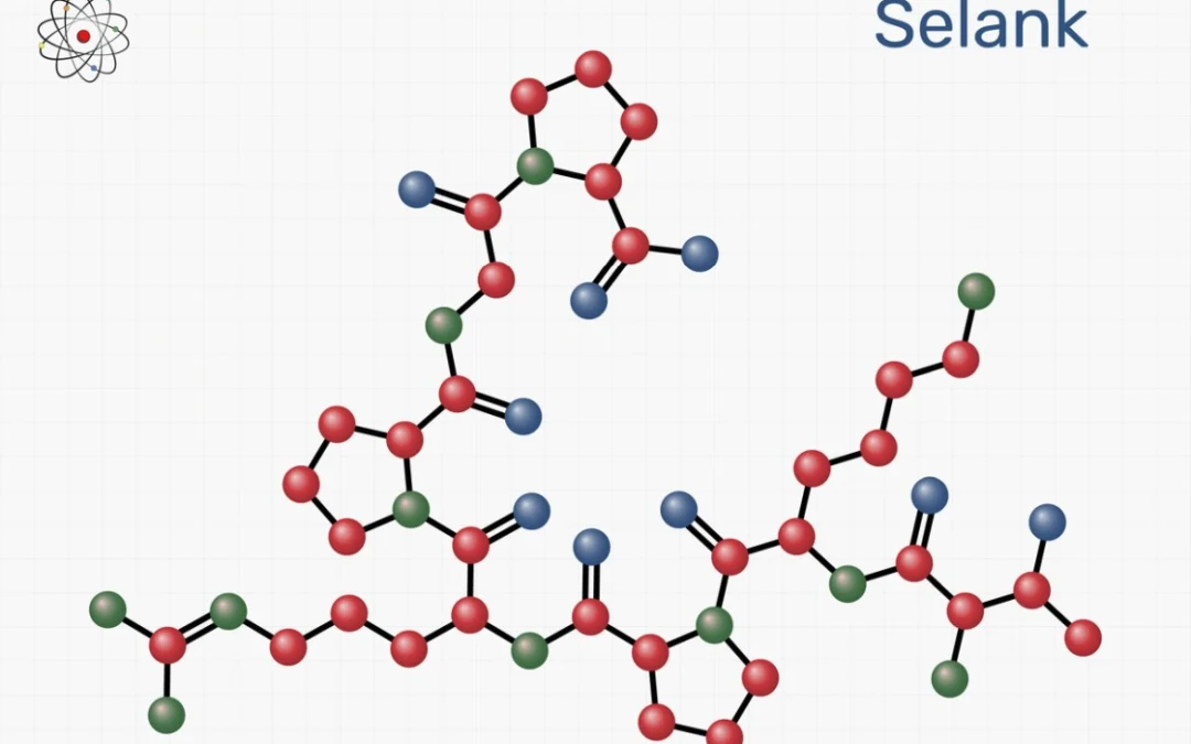
BPC-157 & TB-500 Blend: An Exploration in Cell Proliferation and Regeneration

BPC-157 & TB-500 Blend: An Exploration in Cell Proliferation and Regeneration

The BPC-157 & TB-500 blend incorporates two synthetic peptides with very different structures but similar research potential. TB-500 is a synthetic version of the thymosin beta-4 (Tβ4) peptide that naturally occurs in the cells of the thymus gland. It is made of 43 amino acids encoded by the TMSB4X gene. The peptide has the following sequence: SDKPDMAEI EKFDKSKLKK TETQEKNPLP SKETIEQEKQ AGES, and its molecular weight is 4921 g/mol. Based on thymosin beta-4 research, TB-500 may regulate cell migration, differentiation, and tissue repair.[1] It is thought to interact with different signaling pathways within cells to exert its action. Studies also suggest that TB-500 may potentially promote angiogenesis, which is the formation of new blood vessels. Additionally, it has been suggested by experimental models that TB-500 might contribute to cellular and tissue regeneration.

BPC-157, also known as L 14736, PL-10, and bepecin, is a much shorter peptide comprising 15 amino acids. This pentadecapeptide is fully synthetic, but its structure is derived from the biologically active sequence of another peptide naturally found in gastric juices. BPC-157 may be associated with various cellular processes and is believed to play a role in tissue repair. It is thought to interact with signaling pathways within cells, potentially influencing factors involved in regeneration and recovery. Some studies suggest that BPC-157 may also have the ability to promote angiogenesis and modulate inflammatory processes.[2] Additionally, it has been suggested by experimental models that BPC-157 might contribute to the protection and regeneration of different cells and tissues.


BPC-157 & TB-500 Blend Mechanisms of Action

BPC-157 & TB-500 appear to have different mechanisms, despite their similar potential. For example, TB-500 (Tβ4) appears to regulate the cellular actin-cytoskeleton and cellular migration by sequestering G-actin.[3] [4] Furthermore, studies suggest that there appears to be a specific signaling pathway involved with damaged tissues that TB-500 may regulate. For instance, researchers have conducted experiments using cell models and observed that TB-500 potentially increases the expression of microRNA-146a (miR-146a), which might be a negative regulator of specific signaling pathways in cells. It appeared that this resulted in TB-500 possibly decreasing the expression of two proinflammatory cytokines associated with the aforementioned signaling pathways – L-1 receptor-associated kinase 1 (IRAK1) and tumor necrosis factor receptor-associated factor 6 (TRAF6). As a further suggestion that the increased expression of miR-146a may be TB-500’s main mechanism of action, the researchers commented that “transfection of anti-miR-146a nucleotides reversed the inhibitory effect of Tβ4 on IRAK1 and TRAF6.[5]  

On the other hand, BPC-157 appears to exert its actions via multiple mechanisms, which may include nitric oxide synthesis, regulating cells involved in tissue repair, growth factors, and inflammation. There is a possibility that BPC-157 interacts with the NO system, potentially offering protection to the endothelium and possibly inducing angiogenic action by promoting the formation of new blood vessels.[6] It may have the potential to stimulate the expression of the early growth response 1 gene, which might be responsible for generating cytokines and growth factors, and perhaps facilitating the early formation of the extracellular matrix, including collagen. It should be noted that BPC-157’s interaction with nerve growth factor 1-A binding protein-2 might have repressive effects on certain factors. However, further research is needed to fully understand and confirm these potential mechanisms of BPC-157.


BPC-157 & TB-500 Blend and the Musculoskeletal System

The BPC-157 & TB-500 appear to both have the potential for speeding up the regeneration of connective tissue, such as the one found in tendons and ligaments. In one murine study, TB-500 was investigated for its potential on ligament recovery and regeneration.[7] The scientists performed histological analysis to compare TB-500 against a placebo in a model of ligament injury. They commended that TB-500 may have induced the formation of a more uniform and evenly spaced bundles of collagen fibers within the granulation tissue that also have larger diameters compared to the control. Furthermore, the mechanical properties of the regenerating tissues, including the femur-medial collateral ligament-tibia complexes, appeared to be improved in the TB-500 group compared to the control.

Another study explored the potential impact of BPC 157 on the outgrowth of tendon fibroblasts from cultured tendon explants.[8] The findings suggested that BPC 157 possibly enhanced the outgrowth of tendon explants. BPC 157 potentially increased the survival of these cells under H(2)O(2) stress. Furthermore, the researchers commented that “BPC 157 markedly increased the in vitro migration of tendon fibroblasts in a dose-dependent manner as revealed by transwell filter migration assay. BPC 157 also dose-dependently accelerated the spreading of tendon fibroblasts on culture dishes.” This effect was potentially associated with the induction of F-actin formation, as evidenced by FITC-phalloidin staining. The study also investigated the potential involvement of the FAK-paxillin (two focal adhesion–associated proteins that transmit signals downstream of integrins) pathway in mediating the potential of BPC 157. Western blot analysis suggested that the phosphorylation levels of both FAK and paxillin were apparently increased by BPC 157, while the total amounts of protein remained unchanged.


Conclusion

In summary, the BPC-157 & TB-500 blend shows a synergistic research potential. TB-500 may be involved in regulating cell migration, differentiation, and repair, potentially promoting angiogenesis and connective tissue regeneration. Furthermore, BPC-157 is also believed to have a potential in cellular repair, possibly by influencing factors involved in the normal recovery process. These peptides appear to have different mechanisms of action, with TB-500 possibly regulating cellular actin-cytoskeleton and migration, while BPC-157 may interact with various processes such as nitric oxide synthesis, growth factors, and inflammation regulation. Both peptides show potential in speeding up the regeneration of connective tissue, such as tendons and ligaments, with TB-500 potentially improving collagen fiber formation and regeneration and BPC-157 potentially enhancing tendon fibroblast outgrowth and migration. Further research is needed to confirm these mechanisms.

Disclaimer: The products mentioned are not intended for human or animal consumption. Research chemicals are intended solely for laboratory experimentation and/or in-vitro testing.  Bodily introduction of any sort is strictly prohibited by law.  All purchases are limited to licensed researchers and/or qualified professionals. All information shared in this article is for educational purposes only.


References


  1. Maar, K., Hetenyi, R., Maar, S., Faskerti, G., Hanna, D., Lippai, B., Takatsy, A., & Bock-Marquette, I. (2021). Utilizing Developmentally Essential Secreted Peptides Such as Thymosin Beta-4 to Remind the Adult Organs of Their Embryonic State-New Directions in Anti-Aging Regenerative Therapies. Cells, 10(6), 1343. https://doi.org/10.3390/cells10061343
  2. Seiwerth, S., Milavic, M., Vukojevic, J., Gojkovic, S., Krezic, I., Vuletic, L. B., Pavlov, K. H., Petrovic, A., Sikiric, S., Vranes, H., Prtoric, A., Zizek, H., Durasin, T., Dobric, I., Staresinic, M., Strbe, S., Knezevic, M., Sola, M., Kokot, A., Sever, M., … Sikiric, P. (2021). Stable Gastric Pentadecapeptide BPC 157 and Wound Healing. Frontiers in pharmacology, 12, 627533. https://doi.org/10.3389/fphar.2021.627533
  3. Huff, T., Müller, C. S., Otto, A. M., Netzker, R., & Hannappel, E. (2001). beta-Thymosins, small acidic peptides with multiple functions. The international journal of biochemistry & cell biology, 33(3), 205–220. https://doi.org/10.1016/s1357-2725(00)00087-x
  4. Sanders, M. C., Goldstein, A. L., & Wang, Y. L. (1992). Thymosin beta 4 (Fx peptide) is a potent regulator of actin polymerization in living cells. Proceedings of the National Academy of Sciences of the United States of America, 89(10), 4678–4682. https://doi.org/10.1073/pnas.89.10.4678
  5. Santra, M., Zhang, Z. G., Yang, J., Santra, S., Santra, S., Chopp, M., & Morris, D. C. (2014). Thymosin β4 up-regulation of microRNA-146a promotes oligodendrocyte differentiation and suppression of the Toll-like proinflammatory pathway. The Journal of biological chemistry, 289(28), 19508–19518. https://doi.org/10.1074/jbc.M113.529966
  6. Sikiric, P., Seiwerth, S., Rucman, R., Turkovic, B., Rokotov, D. S., Brcic, L., Sever, M., Klicek, R., Radic, B., Drmic, D., Ilic, S., Kolenc, D., Stambolija, V., Zoricic, Z., Vrcic, H., & Sebecic, B. (2012). Focus on ulcerative colitis: stable gastric pentadecapeptide BPC 157. Current medicinal chemistry, 19(1), 126–132. https://doi.org/10.2174/092986712803414015
  7. Xu, B., Yang, M., Li, Z., Zhang, Y., Jiang, Z., Guan, S., & Jiang, D. (2013). Thymosin β4 enhances the healing of medial collateral ligament injury in rat. Regulatory peptides, 184, 1–5. https://doi.org/10.1016/j.regpep.2013.03.026
  8. Chang, C. H., Tsai, W. C., Lin, M. S., Hsu, Y. H., & Pang, J. H. (2011). The promoting effect of pentadecapeptide BPC 157 on tendon healing involves tendon outgrowth, cell survival, and cell migration. Journal of applied physiology (Bethesda, Md. : 1985), 110(3), 774–780. https://doi.org/10.1152/japplphysiol.00945.2010
The Neuroendocrine Potential of Hexarelin

The Neuroendocrine Potential of Hexarelin

Hexarelin is a synthetic hexapeptide that belongs to a class of growth hormone-releasing peptides (GHRPs). It is also known as examorelin, EP-23905, and MF-6003. The amino acid sequence of hexarelin is His – D-2-methyl – Trp – Ala – Trp – D-Phe – Lys -NH2. This hexapeptide was derived from GHRP-6 (growth hormone-releasing peptide 6), and it appears to share no sequence similarity with ghrelin. However, researchers posit that it may exhibit an agonistic potential towards the ghrelin receptor and, therefore, may mimic the actions of ghrelin.

Hexarelin studies suggest its high selectivity for the ghrelin receptor, also known as the growth hormone secretagogue receptor 1a (GHSR1a). It appears to exhibit potent activity in stimulating growth hormone release, causing researchers to classify it as a growth hormone secretagogue. Its ability to selectively activate the GHSR1a sets it apart from other compounds in this class. Furthermore, Hexarelin is a unique peptide that appears to be resistant to degradation by digestive enzymes. Yet, the precise mechanisms of action and the full range of Hexarelin’s potential are not definitively established, and its specific interactions with growth hormone secretagogue receptors are still the subject of ongoing research.


Hexarelin Peptide and the Central Nervous System

Hexarelin, a peptide compound, appears to interact with the GHSR1a in the central nervous system, potentially initiating a cascade of intracellular signaling events that modulate various physiological processes. This interaction is hypothesized to occur due to the high affinity and specificity between Hexarelin and the binding site of GHSR1a receptors.

Research studies suggest that as Hexarelin binds to GHSR1a receptors, it may activate intracellular signaling pathways. These pathways are considered to involve molecular events, including the activation of G-proteins like Gαq/11, leading to the activation of phospholipase C (PLC). PLC induces the hydrolysis of phosphatidylinositol 4,5-bisphosphate (PIP2) into inositol triphosphate (IP3) and diacylglycerol (DAG). IP3 release may initiate the mobilization of intracellular calcium stores, potentially increasing cytosolic calcium levels.[1] [2] This rise in calcium concentration may regulate cellular processes such as neurotransmitter release, gene expression, and intracellular signaling cascades. Additionally, DAG may activate protein kinase C (PKC), phosphorylating target proteins and potentially regulating cellular functions.

Activation of GHSR1a receptors by Hexarelin may lead to the release of specific hormonal substances from the pituitary gland. These potential substances include growth hormone (GH), prolactin, adrenocorticotropic hormone (ACTH), and cortisol.[3] [4] Furthermore, the interaction between Hexarelin and GHSR1a receptors in the hippocampus potentially impacts neuronal function and communication. Its specific impact on cellular processes in the hippocampus is not fully understood but is suggested to play a role in the modulation of synaptic plasticity, neurotransmitter release, and mechanisms involved in memory consolidation and cognitive processes.

Furthermore, one study investigated the protective potential of Hexarelin peptide on the central nervous system, particularly the hippocampus. The study used a rat model of neonatal hypoxia-ischemia induced by carotid ligation and hypoxic exposure.[5] The researchers commented that “damage was reduced by 39% in the treatment group, compared with the vehicle group, and injury was significantly reduced in the cerebral cortex, hippocampus, and thalamus but not in the striatum.” Moreover, the study reported that reduction in brain damage appeared to coincide with a decrease in caspase-3 activity, an enzyme involved in cell death, and an increase in the phosphorylation of Akt and glycogen synthase kinase-3beta. Overall, the activation of the Akt signaling pathway appeared to play a role in reducing cell death by modulating glycogen synthase kinase-3beta activity and possibly inhibiting caspase-dependent cell death. 


Hexarelin Peptide and the Ghrelin (GHSR1a) Receptors

Hexarelin peptide has complex potential interactions with the ghrelin receptors in the pituitary gland and the hypothalamus. Moreover, these interactions may be modified by other hormonal substances. For example, certain androgens may have the potential to upregulate ghrelin receptors, which could potentially enhance the response of these receptors to Hexarelin.[6] It is hypothesized that androgens might modulate the expression or sensitivity of ghrelin receptors at the cellular level, although the precise mechanisms behind this interaction are not fully understood. It is possible that androgens could alter gene expression or intracellular signaling pathways, increasing the expression or sensitivity of ghrelin receptors. If this modulation occurs, it is speculated that Hexarelin may have a greater binding affinity to these upregulated receptors, potentially resulting in an amplified Hexarelin action and an apparent release of growth hormone.[7]

Another factor that may potentially enhance the interaction between Hexarelin and the GHSR1a is the simultaneous activation of another receptor found on the pituitary gland, the growth hormone-releasing hormone (GHRH) receptor. For example, researchers have simultaneously tested Hexarelin (HEX) and the natural GHRH-receptor activator – GHRH.[8] They commented that “the two substances induced a true synergistic effect, with GH release after HEX plus GHRH (…) being higher (…) than the arithmetic sum of the GH increases induced by each compound separately.” Other studies have also commented on similar findings.[9] [10] This apparent synergism may be due to two potential mechanisms. Firstly, Hexarelin may potentially sensitize somatotroph cells in the pituitary gland, making them more responsive to the stimulatory action of GHRH. This increased sensitivity may lead to a higher potential for GH release when Hexarelin and GHRH are used together compared to using either compound alone. Secondly, both Hexarelin and GHRH may inhibit the release of somatostatin, a hormone that is considered to suppress GH secretion. By acting together, Hexarelin and GHRH may express a higher potential for somatostatin suppression, allowing for increased GH secretion.


Conclusion

In conclusion, Hexarelin peptide may have potential interactions with the ghrelin receptor (GHSR1a) in the central nervous system. Its proposed resistance to degradation by digestive enzymes further distinguishes it from other compounds in its class. It may modulate intracellular signaling pathways and stimulate growth hormone release from pituitary cells. Furthermore, it may affect the release of specific hormonal substances and appears to play a role in synaptic plasticity, memory consolidation, and cognitive processes. Hexarelin’s interactions with other hormonal substances, such as androgens and growth hormone-releasing hormone (GHRH), may influence its action. 

Disclaimer: The products mentioned are not intended for human or animal consumption. Research chemicals are intended solely for laboratory experimentation and/or in-vitro testing.  Bodily introduction of any sort is strictly prohibited by law.  All purchases are limited to licensed researchers and/or qualified professionals. All information shared in this article is for educational purposes only.


References


  1. Khatib, N., Gaidhane, S., Gaidhane, A. M., Khatib, M., Simkhada, P., Gode, D., & Zahiruddin, Q. S. (2014). Ghrelin: ghrelin as a regulatory Peptide in growth hormone secretion. Journal of clinical and diagnostic research : JCDR, 8(8), MC13–MC17. https://doi.org/10.7860/JCDR/2014/9863.4767
  2. Yin, Y., Li, Y., & Zhang, W. (2014). The growth hormone secretagogue receptor: its intracellular signaling and regulation. International journal of molecular sciences, 15(3), 4837–4855. https://doi.org/10.3390/ijms15034837
  3. Frieboes, R. M., Antonijevic, I. A., Held, K., Murck, H., Pollmächer, T., Uhr, M., & Steiger, A. (2004). Hexarelin decreases slow-wave sleep and stimulates the secretion of GH, ACTH, cortisol and prolactin during sleep in healthy volunteers. Psychoneuroendocrinology, 29(7), 851–860. https://doi.org/10.1016/S0306-4530(03)00152-5
  4. Imbimbo, B. P., Mant, T., Edwards, M., Amin, D., Dalton, N., Boutignon, F., Lenaerts, V., Wüthrich, P., & Deghenghi, R. (1994). Growth hormone-releasing activity of hexarelin in humans. A dose-response study. European journal of clinical pharmacology, 46(5), 421–425. https://doi.org/10.1007/BF00191904
  5. Brywe, K. G., Leverin, A. L., Gustavsson, M., Mallard, C., Granata, R., Destefanis, S., Volante, M., Hagberg, H., Ghigo, E., & Isgaard, J. (2005). Growth hormone-releasing peptide hexarelin reduces neonatal brain injury and alters Akt/glycogen synthase kinase-3beta phosphorylation. Endocrinology, 146(11), 4665–4672. https://doi.org/10.1210/en.2005-0389
  6. Loche, S., Cambiaso, P., Carta, D., Setzu, S., Imbimbo, B. P., Borrelli, P., Pintor, C., & Cappa, M. (1995). The growth hormone-releasing activity of hexarelin, a new synthetic hexapeptide, in short normal and obese children and in hypopituitary subjects. The Journal of clinical endocrinology and metabolism, 80(2), 674–678. https://doi.org/10.1210/jcem.80.2.7852535
  7. Loche, S., Colao, A., Cappa, M., Bellone, J., Aimaretti, G., Farello, G., Faedda, A., Lombardi, G., Deghenghi, R., & Ghigo, E. (1997). The growth hormone response to hexarelin in children: reproducibility and effect of sex steroids. The Journal of clinical endocrinology and metabolism, 82(3), 861–864. https://doi.org/10.1210/jcem.82.3.3795
  8. Arvat, E., Di Vito, L., Gianotti, L., Ramunni, J., Boghen, M. F., Deghenghi, R., Camanni, F., & Ghigo, E. (1997). Mechanisms underlying the negative growth hormone (GH) autofeedback on the GH-releasing effect of hexarelin in man. Metabolism: clinical and experimental, 46(1), 83–88. https://doi.org/10.1016/s0026-0495(97)90173-6
  9. Massoud, A. F., Hindmarsh, P. C., & Brook, C. G. (1996). Hexarelin-induced growth hormone, cortisol, and prolactin release: a dose-response study. The Journal of clinical endocrinology and metabolism, 81(12), 4338–4341. https://doi.org/10.1210/jcem.81.12.8954038
  10. Arvat, E., Gianotti, L., Di Vito, L., Imbimbo, B. P., Lenaerts, V., Deghenghi, R., Camanni, F., & Ghigo, E. (1995). Modulation of growth hormone-releasing activity of hexarelin in man. Neuroendocrinology, 61(1), 51–56. https://doi.org/10.1159/000126827
CJC-1295 & Ipamorelin & GHRP-2 Blend and Pituitary Signaling

CJC-1295 & Ipamorelin & GHRP-2 Blend and Pituitary Signaling

CJC-1295 & Ipamorelin & GHRP-2 are peptides that appear to have an affinity for receptors in the central nervous system. More specifically, they may interact with the pituitary gland cells and the hypothalamus. They are hypothesized to exert neuroendocrine actions, and their potential is tightly linked to their distinctive structures.

CJC-1295 belongs to a class of molecules known as growth hormone-releasing hormone (GHRH) agonists. It is a tetrasubstituted version of GHRH 1-29, representing the shortest functional sequence of GHRH. GHRH 1-29 consists of the first 29 amino acids of the native GHRH peptide and may potentially stimulate growth hormone production in pituitary gland cells. CJC-1295 is also modified by adding a drug affinity complex (DAC) component that may bind to plasma proteins. More specifically, the DAC component in CJC-1295 refers to the attachment of the N-epsilon-3-maleimidopropionamide derivative of lysine at the C terminus. By combining the tetrasubstituted amino acid sequence and the DAC component, CJC-1295 appears to exhibit improved pharmacokinetics while having a similar affinity to the GHRH receptors in the pituitary gland as native GHRH. More specifically, researchers comment that when the peptide was “selected for further pharmacokinetic evaluation, where it was found to be present in plasma beyond 72 h.[1] 

Ipamorelin is a synthetic pentapeptide that is considered to bind to another receptor found in pituitary gland cells, called growth hormone secretagogue receptor (GHS-R1a). These receptors are also found in the hypothalamus. Furthermore, GHS-R1a are also known as the ghrelin receptors, as ghrelin appears to be their main natural ligand. GHRP-2, or Growth Hormone Releasing Peptide 2, is a synthetic peptide composed of six amino acids. It also appears to bind to the ghrelin (GHS-R1a) receptors. By activating them, both Ipamorelin & GHRP-2 appear to stimulate the production of growth hormones in pituitary cells.


CJC-1295 & Ipamorelin & GHRP-2 and the GHRH Receptor

CJC-1295 & Ipamorelin & GHRP-2 may interact with various receptors found in the cells of the pituitary gland, such as the GHRH receptor. More specifically, the GHRH receptor is likely the primary target of CJC-1295. 

When the peptide potentially interacts with the growth hormone-releasing hormone GHRH receptor, it appears to bind with specific binding sites on the receptor protein, which may lead to conformational changes in the receptor structure. This binding event may initiate a cascade of molecular events that appear to activate signal transduction pathways within the target cells. 

The apparent conformational changes induced by CJC-1295 binding facilitate the potential activation of G-proteins, which are intracellular signaling proteins that may act as molecular switches. These G-proteins appear to be associated with the intracellular side of the GHRH receptor.[2] Activated G-proteins may stimulate the production of second messengers like cyclic adenosine monophosphate (cAMP) or inositol trisphosphate (IP3), which may serve as secondary signaling molecules, propagating the signal further into the cell.[3]

Second messengers such as cAMP may also activate protein kinases, which appear to be enzymes responsible for phosphorylating specific target proteins. Protein kinases may play a role in regulating various cellular processes. Activation of protein kinases might lead to the phosphorylation of transcription factors, which are proteins potentially involved in controlling gene expression. Once phosphorylated, these transcription factors may enter the nucleus and potentially modulate the transcription of specific genes associated with growth hormone synthesis and secretion. Ultimately, the molecular events triggered by CJC-1295 binding may result in the fusion of secretory vesicles containing growth hormone with the plasma membrane. This fusion enables the potential release of growth hormone outside the pituitary cells, where it may exert biological action.[4]


CJC-1295 & Ipamorelin & GHRP-2 and the GHS-R1a

The GHS-R1a receptors are found in the pituitary gland and the hypothalamus and appear to be the main targets of Ipamorelin & GHRP-2. To understand their molecular mechanisms of interaction with GHS-R1a, it’s important to delve into the structure and function of both the receptor and the peptides. 

GHS-R1a is a G-protein coupled receptor (GPCR) that belongs to the class A rhodopsin-like family of receptors. It appears to play a potential role in regulating growth hormone release by pituitary cells into the extracellular environment.[5] Scientists comment, “since its discovery, hundreds of studies have shown the importance of this receptor and its endogenous ligand, ghrelin, in metabolism, neurotransmission, and behavior.” The receptor consists of seven transmembrane domains, an extracellular N-terminus, and an intracellular C-terminus. The extracellular N-terminus of GHS-R1a may have the potential for ligand binding to agonists such as Ipamorelin & GHRP-2, while the intracellular C-terminus interacts with G-proteins to initiate signaling pathways.[6]

Both Ipamorelin and GHRP-2 may act as agonists for GHS-R1a, meaning they potentially bind to the receptor and activate its signaling pathway. These peptides have a specific sequence of amino acids that allows them to potentially interact with GHS-R1a. The N-terminus of GHS-R1a contains binding sites that appear to recognize specific amino acid sequences in Ipamorelin and GHRP-2. When these peptides come into contact with the receptor, they appear to bind to these sites through non-covalent interactions such as hydrogen bonds, electrostatic interactions, and van der Waals forces. Upon Ipamorelin or GHRP-2 potentially binding to GHS-R1a, there appears to be a conformational change in the receptor. This change potentially leads to activating intracellular signaling pathways, primarily involving the G-proteins. GHS-R1a may interact with G-proteins, specifically the Gαq/11 subunit.[7]

Activation of Gαq/11 triggers the release of a signaling molecule called GTP (guanosine triphosphate) from GDP (guanosine diphosphate) bound to Gαq/11. The GTP-bound Gαq/11 subunit dissociates from the receptor and may activate downstream signaling events. Furthermore, the dissociation of Gαq/11 from GHS-R1a may initiate a series of intracellular signaling cascades. 

One of the primary signaling pathways potentially activated by GHS-R1a involves the enzyme phospholipase C (PLC). Gαq/11 binds to PLC, which in turn may cleave a phospholipid called phosphatidylinositol 4,5-bisphosphate (PIP2) into two secondary messengers: inositol trisphosphate (IP3) and diacylglycerol (DAG).[8] IP3 appears to bind to receptors on the endoplasmic reticulum, causing the release of calcium ions (Ca2+) from intracellular stores. Furthermore, DAG may activate protein kinase C (PKC), phosphorylating downstream signaling molecules, further amplifying the signaling cascade. These signaling events potentially lead to the activation of protein kinases, transcription factors, and other regulatory proteins involved in the growth hormone release by pituitary cells into the extracellular environment.


Conclusion

In conclusion, CJC-1295 & Ipamorelin & GHRP-2 may interact with receptors in the central nervous system, particularly in the pituitary gland cells and hypothalamus. These peptides are hypothesized to have neuroendocrine actions, and their unique structures are directly linked to their secretagogue-like potential. CJC-1295 may bind to GHRH receptors in the pituitary gland, potentially leading to conformational changes and the activation of intracellular signaling pathways for growth hormone production. Ipamorelin and GHRP-2 may bind to GHS-R1a receptors, also known as ghrelin receptors which may initiate a cascade of molecular events involving G-proteins, second messengers, protein kinases, and transcription factors, potentially resulting in the release of growth hormone. 

Disclaimer: The products mentioned are not intended for human or animal consumption. Research chemicals are intended solely for laboratory experimentation and/or in-vitro testing. Bodily introduction of any sort is strictly prohibited by law. All purchases are limited to licensed researchers and/or qualified professionals. All information shared in this article is for educational purposes only.


References


  1. Jetté, L., Léger, R., Thibaudeau, K., Benquet, C., Robitaille, M., Pellerin, I., Paradis, V., van Wyk, P., Pham, K., & Bridon, D. P. (2005). Human growth hormone-releasing factor (hGRF)1-29-albumin bioconjugates activate the GRF receptor on the anterior pituitary in rats: identification of CJC-1295 as a long-lasting GRF analog. Endocrinology, 146(7), 3052–3058. https://doi.org/10.1210/en.2004-1286
  2. Martin, B., Lopez de Maturana, R., Brenneman, R., Walent, T., Mattson, M. P., & Maudsley, S. (2005). Class II G protein-coupled receptors and their ligands in neuronal function and protection. Neuromolecular medicine, 7(1-2), 3–36. https://doi.org/10.1385/nmm:7:1-2:003
  3. Newton, A. C., Bootman, M. D., & Scott, J. D. (2016). Second Messengers. Cold Spring Harbor perspectives in biology, 8(8), a005926. https://doi.org/10.1101/cshperspect.a005926
  4. Ionescu, M., & Frohman, L. A. (2006). Pulsatile secretion of growth hormone (GH) persists during continuous stimulation by CJC-1295, a long-acting GH-releasing hormone analog. The Journal of clinical endocrinology and metabolism, 91(12), 4792–4797. https://doi.org/10.1210/jc.2006-1702
  5. Albarrán-Zeckler, R. G., & Smith, R. G. (2013). The ghrelin receptors (GHS-R1a and GHS-R1b). Endocrine development, 25, 5–15. https://doi.org/10.1159/000346042
  6. Childs, M. D., & Luyt, L. G. (2020). A Decade’s Progress in the Development of Molecular Imaging Agents Targeting the Growth Hormone Secretagogue Receptor. Molecular imaging, 19, 1536012120952623. https://doi.org/10.1177/1536012120952623
  7. Yin, Y., Li, Y., & Zhang, W. (2014). The growth hormone secretagogue receptor: its intracellular signaling and regulation. International journal of molecular sciences, 15(3), 4837–4855. https://doi.org/10.3390/ijms15034837
  8. Bill, C. A., & Vines, C. M. (2020). Phospholipase C. Advances in experimental medicine and biology, 1131, 215–242. https://doi.org/10.1007/978-3-030-12457-1_9
NAD+ Peptide and Cellular Aging

NAD+ Peptide and Cellular Aging

NAD+ peptide (nicotinamide adenine dinucleotide) is a coenzyme that plays a potential role in various biological processes, particularly in cellular metabolism. It is derived from the B-vitamin niacin (vitamin B3) in redox reactions, transferring electrons during metabolic reactions.

NAD+ exists in two forms: oxidized (NAD+) and reduced (NADH). The scientific theory posits that the oxidized form, NAD+, accepts electrons during reactions, while the reduced form, NADH, donates electrons. This interconversion between NAD+ and NADH may allow for the transfer of energy and electrons between molecules in metabolic pathways.

Nicotinamide Adenine Dinucleotide appears to participate in several essential metabolic processes, including cellular respiration, DNA repair, redox reactions, and metabolic regulation. NAD+ may be a crucial player in glycolysis, the Krebs cycle (citric acid cycle), and oxidative phosphorylation. During these processes, NAD+ may accept electrons and become reduced to NADH, possibly carrying the electrons to the electron transport chain for ATP production. NAD+ appears also to be involved in regulating several other metabolic pathways, including lipid metabolism and the biosynthesis of macromolecules.

Furthermore, NAD+ may be involved in DNA repair mechanisms, particularly in a class of enzymes called sirtuins. Sirtuins are considered to utilize NAD+ as a cofactor to remove acetyl groups from proteins, possibly regulating their activity and influencing DNA repair, gene expression, and cellular longevity.


NAD+ Peptide and Cellular Longevity

Researchers posit NAD+’s potential in various processes related to cellular longevity, particularly in DNA repair and gene expression regulation through sirtuins.[1] Sirtuins are a class of proteins that are considered to require NAD+ as a cofactor to carry out possible enzymatic activities. They are potentially involved in various cellular processes, including DNA repair, gene expression, and metabolic regulation. They have been linked to cellular longevity and lifespan.[2] The Sirtuin family of enzymes, such as SIRT1, appear to be NAD+-dependent protein deacetylases and may have the potential to sense NAD+ levels. Scientific reviews hypothesize that sirtuins may primarily sense NAD+ not NADH or the NAD+/NADH ratio.[3] However, the mechanisms by which enzymes like sirtuins respond to cell redox changes are not fully understood.

Researchers have proposed that the decline in NAD+ levels that may occur with aging might contribute to age-related cellular dysfunction, and restoring or maintaining NAD+ levels might potentially mitigate it. For example, one experiment in worms suggested that NAD(+) levels decline with age and that reducing NAD(+) may further decrease lifespan.[4] Conversely, restoring NAD(+) through genetic or other means appeared to promote longevity in worms. The researchers hypothesized that these actions might rely on the activity of the SIRT1, otherwise known as Protein Deacetylase Sir-2.1 or Silent Information Regulator 2.1, and may involve the induction of imbalances in mitochondrial proteins, as well as the activation of stress signaling pathways like the mitochondrial unfolded protein response (UPR(mt)) and the nuclear translocation and activation of the FOXO transcription factor DAF-16. The authors also considered that enhancing mitochondrial stress signaling by modulating NAD(+) levels might be a potential strategy to support mitochondrial function.


NAD+ Peptide and Cellular Respiration

Researchers should note that “Nicotinamide adenine dinucleotide (…) is a coenzyme for redox reactions, making it central to energy metabolism.[5] NAD+ may play a crucial role in cellular respiration, the process by which cells convert nutrients into usable energy. Cellular respiration is considered to occur in multiple steps involving different metabolic pathways, and NAD+ has been posited to be involved in two specific stages: glycolysis and the citric acid cycle (also known as the Krebs cycle or tricarboxylic acid/TCA cycle).[6]

Scientists consider that glycolysis, the process by which glucose is broken down into pyruvate, may generate a small amount of ATP (adenosine triphosphate) and NADH. Researchers posit that Nicotinamide Adenine Dinucleotide accepts electrons and a hydrogen ion (H+) from glucose, forming NADH. NADH may carry these high-energy electrons to the next stage of cellular respiration. In the citric acid cycle, the pyruvate produced in glycolysis is considered to break down further, potentially releasing additional energy. NAD+ appears to be involved in several reactions, possibly t accepting electrons and H+ ions, forming NADH. These reactions appear to occur within the mitochondria, the cell’s energy-producing organelles.

NADH that appears to be generated in both glycolysis and the citric acid cycle, has the potential to carry the high-energy electrons to the electron transport chain, the final stage of cellular respiration. Here, NADH may donate its electrons to the chain, allowing them to flow through a series of protein complexes. This electron flow appears to drive protons (H+) pumping across the mitochondrial membrane, potentially establishing an electrochemical gradient. Eventually, the electrons and protons may combine with oxygen to form water, while the energy released by the electron flow may produce ATP through oxidative phosphorylation. As NADH donates its electrons to the electron transport chain, it may be converted back to NAD+, ready to possibly participate in the next round of glycolysis and the citric acid cycle. This regeneration of NAD+ may be instrumental in sustaining possible continuous production of ATP through cellular respiration.


NAD+ Peptide and DNA Repair

One of the key enzymes researched in connection to DNA repair, poly(ADP-ribose) polymerase (PARP), appears to rely on NAD+. When DNA damage occurs, PARP may possibly be activated, potentially bindingto the damaged site on the DNA. PARP may utilize NAD+ as a substrate to add ADP-ribose units to itself and other target proteins, a process termed poly(ADP-ribosylation) by researchers.[7] This modification has the potential to help recruit and activate other DNA repair proteins, possibly facilitating the repair of DNA lesions.

The poly(ADP-ribosylation) process appears to generate poly(ADP-ribose) (PAR) chains, which may serve as a signal for DNA repair machinery to recognize and target damaged DNA. PARP may also play a role in recognizing and repairing single-strand breaks (SSBs) in DNA. Should NAD+ be a cofactor, PARP may help maintain genomic stability by initiating and coordinating DNA repair processes. However, this process may also deplete cellular NAD+ levels, which may impact other NAD+-dependent cellular processes, such as energy metabolism and signaling. Studies comment that “in response to DNA damage, the rate of PAR synthesis increases rapidly up to 500-fold, which can consume a significant amount of NAD+.[8] Thus, the researchers hypothesize that the depletion of intracellular NAD+ due to PARP1 activation may affect the NAD+/SIRT1 axis, possibly leading to defects in mitochondrial homeostasis, ROS (reactive oxygen species) production, DNA repair, and cell survival.


Conclusion

In conclusion, Nicotinamide Adenine Dinucleotide has been suggested by researchers to act as a crucial coenzyme involved in various biological processes, particularly cellular metabolism. It might participate in essential metabolic pathways, such as cellular respiration, DNA repair, and metabolic regulation. NAD+ has been suspected to play a key role in energy production through glycolysis and the citric acid cycle, where researchers suggest it may accept electrons and transfer them to the electron transport chain for ATP synthesis. Additionally, NAD+ appears to be involved in DNA repair mechanisms, specifically through enzymes like sirtuins and PARP, which possibly utilize NAD+ peptide to regulate gene expression, cellular longevity, and genomic stability.

Disclaimer: The products mentioned are not intended for human or animal consumption. Research chemicals are intended solely for laboratory experimentation and/or in-vitro testing.  Bodily introduction of any sort is strictly prohibited by law.  All purchases are limited to licensed researchers and/or qualified professionals. All information shared in this article is for educational purposes only.


References


  1. Imai, S., & Guarente, L. (2014). NAD+ and sirtuins in aging and disease. Trends in cell biology, 24(8), 464–471. https://doi.org/10.1016/j.tcb.2014.04.002
  2. Wątroba, M., Dudek, I., Skoda, M., Stangret, A., Rzodkiewicz, P., & Szukiewicz, D. (2017). Sirtuins, epigenetics and longevity. Aging research reviews, 40, 11–19. https://doi.org/10.1016/j.arr.2017.08.001
  3. Anderson, K. A., Madsen, A. S., Olsen, C. A., & Hirschey, M. D. (2017). Metabolic control by sirtuins and other enzymes that sense NAD+, NADH, or their ratio. Biochimica et biophysica acta. Bioenergetics, 1858(12), 991–998. https://doi.org/10.1016/j.bbabio.2017.09.005
  4. Mouchiroud, L., Houtkooper, R. H., Moullan, N., Katsyuba, E., Ryu, D., Cantó, C., Mottis, A., Jo, Y. S., Viswanathan, M., Schoonjans, K., Guarente, L., & Auwerx, J. (2013). The NAD(+)/Sirtuin Pathway Modulates Longevity through Activation of Mitochondrial UPR and FOXO Signaling. Cell, 154(2), 430–441. https://doi.org/10.1016/j.cell.2013.06.016
  5. Covarrubias, A. J., Perrone, R., Grozio, A., & Verdin, E. (2021). NAD+ metabolism and its roles in cellular processes during ageing. Nature reviews. Molecular cell biology, 22(2), 119–141. target=”_blank” rel=”noopener”https://doi.org/10.1038/s41580-020-00313-x
  6. Ahmad M, Wolberg A, Kahwaji CI. Biochemistry, Electron Transport Chain. [Updated 2022 Sep 5]. In: StatPearls [Internet]. Treasure Island (FL): StatPearls Publishing; 2023 Jan-. Available from: https://www.ncbi.nlm.nih.gov/books/NBK526105/
  7. Leung, A., Todorova, T., Ando, Y., & Chang, P. (2012). Poly(ADP-ribose) regulates post-transcriptional gene regulation in the cytoplasm. RNA biology, 9(5), 542–548. https://doi.org/10.4161/rna.19899
  8. Croteau, D. L., Fang, E. F., Nilsen, H., & Bohr, V. A. (2017). NAD+ in DNA repair and mitochondrial maintenance. Cell cycle (Georgetown, Tex.), 16(6), 491–492. https://doi.org/10.1080/15384101.2017.1285631
N-Acetyl Selank Studies on Cognitive Impact

N-Acetyl Selank Studies on Cognitive Impact

N-Acetyl Selank is an acetylated form of the heptapeptide Selank. Selank is a synthetic analog of the natural tetrapeptide tuftsin (threonine – lysine – proline – arginine). The sequence of N-Acetyl Selank is acetyl – threonine – lysine – proline – arginine – proline – glycine – proline. Adding proline – glycine – proline at the C-terminus and the acetylation at the N-terminus is intended to improve its stability compared to tuftsin. Tuftsin is found in the blood of various mammals as it is normally located in the Fc-domain of the heavy chain of immunoglobulin G (residues 289-292). As such, tuftsin is hypothesized to have immunomodulatory properties.

N-acetyl Selank peptide may potentially influence neurotransmitters, central nervous system signaling, and neuroplasticity. More specifically, the potential of N-acetyl Selank lies in higher cognitive functioning, serotonin signaling, and brain-derived neurotrophic factor (BDNF) expression. Studies on N-acetyl Selank are lacking, but data from Selank’s research may be used as a reference.


N-Acetyl Selank and Higher Cognitive Functions

One murine trial involving trained Winstar rats investigated the potential of the non-acetylated version of N-Acetyl Selank for influencing learning, memory, and serotonin levels.[1] According to the research, N-Acetyl Selank may upregulate serotonin metabolism in the hypothalamus and caudal brain stem for 30 minutes to 2 hours. Additionally, N-Acetyl Selank may have a positive influence on memory trace stability for a period of 30 days. These findings suggest that N-Acetyl Selank may have favorable potential on memory storage processes during the consolidation phase of memory formation. The researchers also speculated that the potential of N-Acetyl Selank for improving higher cognitive functions may be attributed to its possible influence on serotonin levels and metabolites in the brain.

Furthermore, one clinical study by Medvedev et al. (2014) noted the potential of the non-acetylated version of N-Acetyl Selank when used in isolations.[2] The researchers commented on the apparent improvement in the results of several scales for mood, such as HDRS, CGI, Spilberger, and SF-36. They also share that the “effect lasted for a week after last receiving the peptide.

In another publication from 2015, the same researchers also suggest that N-Acetyl Selank may positively influence chemically induced asthenia, attention, and memory impairment.[3] The researchers hypothesized that this might be due to the potential action of N-Acetyl Selank on higher cognitive functions.


N-Acetyl Selank and Enkephalins

Enkephalins are considered to be the natural ligands to the opioid receptors in the nervous system and play a role in mood and nociception. One clinical study suggested that low levels of tau(1/2) leu-enkephalin, a specific parameter related to enkephalin activity in serum, may have anxiogenic potential.[4] Yet, the non-acetylated version of N-Acetyl Selank may potentially increase tau(1/2) leu-enkephalin levels. The researchers also noted that the peptide had an apparently favorable potential on psychometric scales such as Hamilton, Zung, and CGI, likely due to its possible interaction with the enkephalin signaling in the central nervous system.

Another clinical trial posited a decreased half-life of enkephalins and reduced total enkephalinase activity might have anxiogenic activity.[5] The non-acetylated version of N-Acetyl Selank was suggested to block the enzymatic hydrolysis of plasma enkephalins by inhibiting enkephalinase activity. The researchers commented that the peptide “inhibited enzymatic hydrolysis of plasma enkephalin […]. Selank was more potent than peptidase inhibitors bacitracin and puromycin in inhibiting enkephalinases.


N-Acetyl Selank and Serotonin Signaling

Semenova et al. (2009) investigated the potential of the non-acetylated version of N-Acetyl Selank on influencing serotonin (5-HT) signaling in the brains of Wistar rats.[6] The rats were exposed to a 5-HT synthesis inhibitor called p-chlorophenyl alanine (PCPA) four days before the experiment. There were a total of 87 mature rats included in the study. The scientists commented that the non-acetylated version of N-Acetyl Selank may increase 5-HT metabolism in the brain stem despite exposure to PCPA. This suggests that the peptide N-Acetyl Selank may potentially improve disturbances caused by a decrease in 5-HT metabolism.


N-Acetyl Selank and Neurotrophic Growth Factors

Brain-derived neurotrophic factor (BDNF) is a neurotrophic growth factor studied for its potential to support neuroplasticity and synaptogenesis, neural remodeling, neurogenesis, nerve cell growth, survival, and adaptation.[7] [8] Scientists also note, “The neurotrophic factor BDNF is an important regulator for the development of brain circuits, for synaptic and neuronal network plasticity, as well as for neuroregeneration and neuroprotection.[9]

Several researchers have investigated the potential of the non-acetylated version of N-Acetyl Selank on BDNF expression in the rat hippocampus.[10] More specifically, the researchers measured the expression of BDNF mRNA and protein levels, as this neurotrophic growth factor is a potentially important regulator of memory formation.

The reported findings suggest that N-Acetyl Selank may stimulate the expression of BDNF in the hippocampus, indicating a potential role in regulating hippocampal function. This is based on comments that the non-acetylated version of N-Acetyl Selank appeared to upregulate BDNF mRNA expression in the rat hippocampus. Similarly, the protein level of BDNF in the hippocampus also appeared to increase after introducing the peptide. 


Conclusion

In conclusion, N-Acetyl Selank may exhibit the potential to impact neurotransmitters, central nervous system signaling, and neuroplasticity. Studies suggest that N-Acetyl Selank may positively influence higher cognitive functions, including memory and learning processes, potentially through action on serotonin levels and BDNF expression. Furthermore, its proposed interaction with enkephalin signaling in the central nervous system may contribute to its vast research potential.

Disclaimer: The products mentioned are not intended for human or animal consumption. Research chemicals are intended solely for laboratory experimentation and/or in-vitro testing.  Bodily introduction of any sort is strictly prohibited by law.  All purchases are limited to licensed researchers and/or qualified professionals. All information shared in this article is for educational purposes only.


References


  1. Semenova TP, Kozlovskiĭ II, Zakharova NM, Kozlovskaia MM. [Experimental optimization of learning and memory processes by selank]. Eksp Klin Farmakol. 2010 Aug;73(8):2-5. Russian. PMID: 20919548.
  2. Medvedev VE, Tereshchenko ON, Israelian AIu, Chobanu IK, Kost NV, Sokolov OIu, Miasoedov NF. [A comparison of the anxiolytic effect and tolerability of selank and phenazepam in the treatment of anxiety disorders]. Zh Nevrol Psikhiatr Im S S Korsakova. 2014;114(7):17-22. Russian. PMID: 25176261.
  3. Medvedev VE, Tereshchenko ON, Kost NV, Ter-Israelyan AY, Gushanskaya EV, Chobanu IK, Sokolov OY, Myasoedov NF. [Optimization of the treatment of anxiety disorders with selank]. Zh Nevrol Psikhiatr Im S S Korsakova. 2015;115(6):33-40. Russian. doi: 10.17116/jnevro20151156133-40. PMID: 26356395.
  4. Zozulia AA, Neznamov GG, Siuniakov TS, Kost NV, Gabaeva MV, Sokolov OIu, Serebriakova EV, Siranchieva OA, Andriushenko AV, Telesheva ES, Siuniakov SA, Smulevich AB, Miasoedov NF, Seredenin SB. [Efficacy and possible mechanisms of action of a new peptide anxiolytic selank in the therapy of generalized anxiety disorders and neurasthenia]. Zh Nevrol Psikhiatr Im S S Korsakova. 2008;108(4):38-48. Russian. PMID: 18454096.
  5. Zozulya AA, Kost NV, Yu Sokolov O, Gabaeva MV, Grivennikov IA, Andreeva LN, Zolotarev YA, Ivanov SV, Andryushchenko AV, Myasoedov NF, Smulevich AB. The inhibitory effect of Selank on enkephalin-degrading enzymes as a possible mechanism of its anxiolytic activity. Bull Exp Biol Med. 2001 Apr;131(4):315-7. doi: 10.1023/a:1017979514274. PMID: 11550013.
  6. Semenova TP, kozlovskiĭ II, Zakharova NM, Kozlovskaia MM. [Comparison of the effects of selank and tuftsin on the metabolism of serotonin in the brain of rats pretreated with PCPA]. Eksp Klin Farmakol. 2009 Jul-Aug;72(4):6-8. Russian. PMID: 19803361.
  7. Deister C, Schmidt CE. Optimizing neurotrophic factor combinations for neurite outgrowth. J Neural Eng. 2006 Jun;3(2):172-9. doi: 10.1088/1741-2560/3/2/011. Epub 2006 May 16. PMID: 16705273.
  8. Lu B, Figurov A. Role of neurotrophins in synapse development and plasticity. Rev Neurosci. 1997 Jan-Mar;8(1):1-12. doi: 10.1515/revneuro.1997.8.1.1. PMID: 9402641.
  9. Brigadski, T., & Leßmann, V. (2020). The physiology of regulated BDNF release. Cell and tissue research, 382(1), 15–45. https://doi.org/10.1007/s00441-020-03253-2
  10. Inozemtseva LS, Karpenko EA, Dolotov OV, Levitskaya NG, Kamensky AA, Andreeva LA, Grivennikov IA. Intranasal administration of the peptide Selank regulates BDNF expression in the rat hippocampus in vivo. Dokl Biol Sci. 2008 Jul-Aug;421:241-3. doi: 10.1134/s0012496608040066. PMID: 18841804
Acetyl Hexapeptide-3 and Neuromuscular Synapse

Acetyl Hexapeptide-3 and Neuromuscular Synapse

Acetyl Hexapeptide-3 (Argireline) is a synthetic peptide used in scientific research. It is a chain of six amino acids connected in a specific sequence: N-acetyl – L-alpha-glutamyl – L-alpha-glutamyl – L-methionyl – L-glutaminyl – L-arginyl – L-arginine amide. Acetyl Hexapeptide-3 has gained attention for its potential molecular mechanisms that may interact with the release of neurotransmitters in the neuromuscular synapses.

At the molecular level, Acetyl Hexapeptide-3 is hypothesized to act as a competitive inhibitor of SNAP25 (synaptosome-associated protein 25), a component of the SNARE (soluble NSF attachment protein receptor) complex. The protein is named SNAP25 due to its size of 25 kDa. The SNARE complex is considered to play a crucial role in synaptic vesicle Ca(2+)-dependent exocytosis, which involves the release of neurotransmitters such as acetylcholine. Acetylcholine is a neurotransmitter linked to muscle contractions.

By possibly inhibiting acetylcholine exocytosis, Acetyl Hexapeptide-3 may interfere with the function of neuromuscular synapses and prevent the contraction of muscle cells. This mechanism appears similar to the way bacterial toxins, such as the toxin producers by Clostridium botulinum – (BoNTs) – are considered to interact and inhibit neuromuscular synapses. However, Acetyl Hexapeptide-3 may have a potential advantage in terms of permeation through the epidermis due to the attachment of acetyl moiety at the N-terminus of the peptide.[1]

Acetyl Hexapeptide-3 may also interact with other aspects of the function of the nervous system, such as potentially inhibiting nociception. The peptide may also potentially impact collagen production and/or degradation.


Acetyl Hexapeptide-3 and Dermal Topography

Clinical studies suggest that Acetyl Hexapeptide-3 may improve the topography of dermal structures, such as wrinkle depth reduction, by as much as 30% – 48.9%.[2] [3] The scientists posit that the peptide acts by inhibiting the release of neurotransmitters such as acetylcholine in the neuromuscular synapses. They also share that “inhibition of neurotransmitter release was due to the interference of the hexapeptide with the formation and/or stability of the protein complex that is required to drive Ca(2+)-dependent exocytosis, namely the vesicular fusion (known as SNARE) complex.[2]

Researchers also commented that the main potential mechanisms of Acetyl Hexapeptide-3, namely the competitive inhibition of SNAP-25, may be due to sharing a similar amino acid sequence pattern from the N-terminal end of synaptosomal-associated protein-25.[4] This appears to prevent the formation of the ternary soluble N-ethylmaleimide sensitive factor attachment protein receptor (SNARE) complex and interfere with neurotransmitter release, similar to BoNTs. In accordance with the potential action of Acetyl Hexapeptide-3, the contraction of the intrinsic muscles appears to be inhibited.


Acetyl Hexapeptide-3 and Collagen Synthesis

Some studies have investigated the potential Acetyl Hexapeptide-3 influence on collagen synthesis, a vital component of the extracellular matrix considered responsible for the structure and elasticity of connective tissues. The peptide has been suggested to interact with collagen production, potentially affecting the histological structure of dermal tissue.

Murine trials that lasted for at least 6 weeks suggest that the peptide may lead to an apparent increase in type I collagen fibers and a decrease in type III collagen fibers.[5] The scientists used hematoxylin-eosin (HE) and picrosirius-polarization (PSP) stains to assess these histological changes in the dermal tissues. Furthermore, the amount of type I and type III collagen fibers were also semi-quantitatively compared using the software Image-ProPlus.

Due to its potential for reducing type III collagen fibers, Acetyl Hexapeptide-3 may also help reduce scarring during and after tissue regeneration. Some researchers comment that the peptide appears to increase the elasticity of scarred tissue “from 33,5% to 40,5% (…) in left lateral-medial area of the neck and malar area; from 24% to 31,5% (…) in right lateral- medial area of the neck and malar area; and from 25,5% to 38% (…) in forehead and chin area.[6]


Acetyl Hexapeptide-3 and Nociception

Acetyl Hexapeptide-3 has been examined in several in vitro models for its potential impact on various cellular and molecular mechanisms. This includes studying its potential on ion channels and inflammatory processes that are involved in the occurrence of nociceptive problems and hyperalgesia.

For example, studies have investigated the potential of the palmitoylated version of Acetyl Hexapeptide-3, called DD04107, for reducing nociceptive stimuli in models of chronic inflammatory and neuropathic hyperalgesia.[7] More specifically, the research suggests that the palmitoylated version of Acetyl Hexapeptide-3 may inhibit the release of neuromodulators related to nociceptive signaling. It appears to do so by interfering with SNAP-25 and preventing these neuromodulators’ Ca(2+)-dependent exocytosis.As a result, the palmitoylated version of Acetyl Hexapeptide-3 may block the inflammatory recruitment of TRPV1 channels leading to its anti-hyperalgesia and anti-allodynic potential. 

TRPV1 channels are ion channels found predominantly on sensory nerve fibers, including those involved in pain perception. These channels appear to be activated by various stimuli, including heat, inflammatory mediators, and chemical irritants, and their activation is considered to lead to the generation and transmission of pain signals.

In studies using carrageenan-induced inflammation, which helps scientific models to mimic acute inflammatory pain, the palmitoylated version of Acetyl Hexapeptide-3 has been hypothesized to have anti-inflammatory activity by reducing paw volume (a measure of inflammation) and attenuating mechanical hypersensitivity.[8]

Furthermore, the palmitoylated version of Acetyl Hexapeptide-3 has also been evaluated in models of chronic inflammatory pain, such as complete Freund’s adjuvant (CFA)-induced inflammation. In these models, the palmitoylated version of Acetyl Hexapeptide-3 appeared to mitigate thermal hyperalgesia and mechanical allodynia, indicating its potential for chronic inflammatory hyperalgesia.

The palmitoylated version of Acetyl Hexapeptide-3 has also been proposed to potentially reduce pain associated with peripheral neuropathy induced by various agents. 


Conclusion

Acetyl Hexapeptide-3, a synthetic peptide, may have promising potential in various scientific research areas. Its molecular mechanisms involve interacting with neurotransmitter release in neuromuscular synapses by acting as a competitive inhibitor of SNAP25, similar to bacterial toxins. This interference inhibits acetylcholine exocytosis, potentially preventing muscle cell contraction. Furthermore, Acetyl Hexapeptide-3 may impact collagen synthesis, leading to improvements in the topography of dermal structures and a potential reduction in scarring. Studies have also explored its role in nociception, indicating its potential to inhibit neuromodulators related to pain signaling and potentially improve chronic inflammatory and neuropathic hyperalgesia. 

Disclaimer: The products mentioned are not intended for human or animal consumption. Research chemicals are intended solely for laboratory experimentation and/or in-vitro testing.  Bodily introduction of any sort is strictly prohibited by law.  All purchases are limited to licensed researchers and/or qualified professionals. All information shared in this article is for educational purposes only.


References


  1. Grosicki, M., Latacz, G., Szopa, A., Cukier, A., & Kieć-Kononowicz, K. (2014). The study of cellular cytotoxicity of argireline – an anti-aging peptide. Acta biochimica Polonica, 61(1), 29–32.
  2. Blanes-Mira, C., Clemente, J., Jodas, G., Gil, A., Fernández-Ballester, G., Ponsati, B., Gutierrez, L., Pérez-Payá, E., & Ferrer-Montiel, A. (2002). A synthetic hexapeptide (Argireline) with antiwrinkle activity. International journal of cosmetic science, 24(5), 303–310. https://doi.org/10.1046/j.1467-2494.2002.00153.x
  3. Wang, Y., Wang, M., Xiao, X. S., Pan, P., Li, P., & Huo, J. (2013). The anti wrinkle efficacy of synthetic hexapeptide (Argireline) in Chinese Subjects. Journal of cosmetic and laser therapy : official publication of the European Society for Laser Dermatology, Advance online publication. https://doi.org/10.3109/14764172.2012.759234
  4. An, J. H., Lee, H. J., Yoon, M. S., & Kim, D. H. (2019). Anti-Wrinkle Efficacy of Cross-Linked Hyaluronic Acid-Based Microneedle Patch with Acetyl Hexapeptide-8 and Epidermal Growth Factor on Korean Skin. Annals of dermatology, 31(3), 263–271. https://doi.org/10.5021/ad.2019.31.3.263
  5. Wang, Y., Wang, M., Xiao, X. S., Huo, J., & Zhang, W. D. (2013). The anti-wrinkle efficacy of Argireline. Journal of cosmetic and laser therapy : official publication of the European Society for Laser Dermatology, 15(4), 237–241. https://doi.org/10.3109/14764172.2013.769273
  6. Palmieri, B., Noviello, A., Corazzari, V., Garelli, A., & Vadala, M. (2020). Skin scars and wrinkles temporary camouflage in dermatology and oncoesthetics: focus on acetyl hexapeptide-8. La Clinica terapeutica, 171(6), e539–e548. https://doi.org/10.7417/CT.2020.2270
  7. Ponsati, B., Carreño, C., Curto-Reyes, V., Valenzuela, B., Duart, M. J., Van den Nest, W., Cauli, O., Beltran, B., Fernandez, J., Borsini, F., Caprioli, A., Di Serio, S., Veretchy, M., Baamonde, A., Menendez, L., Barros, F., de la Pena, P., Borges, R., Felipo, V., Planells-Cases, R., … Ferrer-Montiel, A. (2012). An inhibitor of neuronal exocytosis (DD04107) displays long-lasting in vivo activity against chronic inflammatory and neuropathic pain. The Journal of pharmacology and experimental therapeutics, 341(3), 634–645. https://doi.org/10.1124/jpet.111.190678
  8. Butrón, D., Zamora-Carreras, H., Devesa, I., Treviño, M. A., Abian, O., Velázquez-Campoy, A., Bonache, M. Á., Lagartera, L., Martín-Martínez, M., González-Rodríguez, S., Baamonde, A., Fernández-Carvajal, A., Ferrer-Montiel, A., Jiménez, M. Á., & González-Muñiz, R. (2021). DD04107-Derived neuronal exocytosis inhibitor peptides: Evidences for synaptotagmin-1 as a putative target. Bioorganic chemistry, 115, 105231. https://doi.org/10.1016/j.bioorg.2021.105231