MGF vs. PEG-MGF: Research in Muscle Regeneration

MGF vs. PEG-MGF: Research in Muscle Regeneration

MGF (Mechano Growth Factor) Mechano growth factor (MGF) is a synthetically produced peptide that researchers suggest may be an alternative splicing variant of insulin-like growth factor 1 (IGF – 1). First described in skeletal muscles, it appears to function as a local tissue repair factor, responding to changes in physiological conditions or environmental stimuli that induce muscle cell proliferation. It is said to activate satellite cells in the muscle and increase the number of desmin positive myogenic precursor cells, potentially leading to hypertrophy or regeneration. Additionally, researchers suggest it may act as a neuroprotective agent in cerebral ischemia. MGF is characterized as IGF – 1Eb with a 52 base pair insert in rats. It appears to have a relatively short half-life of only a few minutes, prompting the development of PEG-MGF to compensate for this disadvantage.

PEG-MGF, or Pegylated Mechano Growth Factor, is a variant of IGF-1. PEG-ylation modifies MGF by fusing polyethylene glycol (PEG) into it, potentially extending its half-life from a few minutes to days, allowing it to travel through the bloodstream for an extended period before breaking down and being excreted by the kidneys. Researchers suggest this process is essential for animal muscle regeneration after physical exertion or injury by promoting nitrogen retention and increasing protein synthesis.


Mechanism of Action

Research data from animal studies suggests that PEG-MGF supports the regeneration process of muscle, positively regulates protein synthesis, and activates satellite cells. In experiments where the PEG-MGF cDNA has been inserted into a plasmid vector and introduced into muscle cells, it appears to be a potent inducer of muscle hypertrophy. The addition of PEG-MGF to muscle myoblasts appeared to have increased proliferation and delayed differentiation, even in the presence of anti – IGF 1 receptor antibodies, potentially by activating fibrinolysis of matrix and metalloproteinase systems. Additionally, researchers propose that intense physical exertion may stimulate the release of growth hormone in the muscles to release MGF, especially as levels of this hormone decrease over time. Furthermore, a recent study of PEG-MGF suggests that the activity of protein kinase C is speculated to be necessary for the activation of this peptide (translocation to the nucleus) of factor 2 related to NFE 2 (Nrf2). This, in turn, is suggested to increase the expression of heme oxygenase 1, an event speculated to mediate neuroprotection of neurons from oxidative stress-induced apoptosis in the brain.


Research into PEG-MGF

In addition to its potential effects on muscle, PEG-MGF is speculated to have other impacts in the bodies of animal research models. Researchers suggest that PEG-MGF may enhance the proliferation and migration of bone marrow-derived mesenchymal stem cells, which are suggested to be a source of autologous stem cells for transplantation to the heart. There is speculative evidence of transient regulation of PEG b MGF expression in response to myocardial infarction associated with ischemia in the heart. Intracoronary exposure to this peptide is suggested to induce myocardial protection and improve hemodynamic function more than mature IGF – 1 after myocardial infarction in sheep. The speculated cellular protection conferred by PEG-MGF is suggested to be based on the inhibition of apoptosis in the border area of the infarct. PEG-MGF has been suggested to stimulate proangiogenic activities in vascular endothelial cells. Therefore, it may confer potentially beneficial action at the level of vascular regeneration and collateralization to restore blood flow to the heart after myocardial infarction. IGF – 1, considered to play an essential role in the interface between neurons in injured or damaged muscles, has shown potential effectiveness in slowing the progression of amyotrophic lateral sclerosis (ALS), a disease characterized by the loss of motor neurons and progressive muscle weakness. Exposure to PEG-MGF has been reported to produce improvements, with more motor neurons surviving in PEG-MGF-treated mice. MGF may be expressed in excess in regenerating regions after global cerebral ischemia. Its transcripts, thought to be expressed during brain development, are suggested to exhibit particular time distributions. Additionally, neonatal hypoxia and insults of hypoxic ischemia are suggested to lead to increased and prolonged expression of only the MGF isoform.


Disclaimer: The products mentioned are not intended for human or animal consumption. Research chemicals are intended solely for laboratory experimentation and/or in-vitro testing.  Bodily introduction of any sort is strictly prohibited by law.  All purchases are limited to licensed researchers and/or qualified professionals. All information shared in this article is for educational purposes only.
Epithalon: Overview of Anti-Aging Experiments

Epithalon: Overview of Anti-Aging Experiments

The science of bioaging is still considered a relatively unexplored territory, with research in biogerontology becoming a focal point in the exploration of anti-cell aging discovery.  To study the effects of a substance that potentially impacts the aging cycle of a cell, Prof. Vladimir Khavinson conducted several methodical research studies on murine models, and developed interesting theories, one of which was the development and exploration of the research peptide Epithalon, which Khavinson et al. suggests may induce anti-aging characteristics upon cells, via telomerase activation and the elongation of telomeres.


What is Epithalon?

Epithalon, also known as Epithalon or Epithalamine, is a synthetic pineal tetrapeptide comprising four amino acids (Alanine, Glutamic acid, Aspartic acid, Glycine). It is considered identical to the natural peptide called Epithalamin, a hormone produced by the pineal gland. The peptide molecule appears to interact with and induce sensitivity in the hypothalamus towards the natural hormone, monitoring the circadian rhythm and controlling melatonin production. Researchers suggest that this peptide may exert positive influences in the realms of cell aging, as well as potentially inhibiting the proliferation of cancer cells, including those in the breast, prostate, and colon. Epithalon is suggested to impact metabolism by increasing the sensitivity of the hypothalamus to its natural hormone influences. This is said to regulate the level of melatonin in the bodies of animal test subjects, thereby regulating the circadian rhythm.


What is Meant by “Aging”?

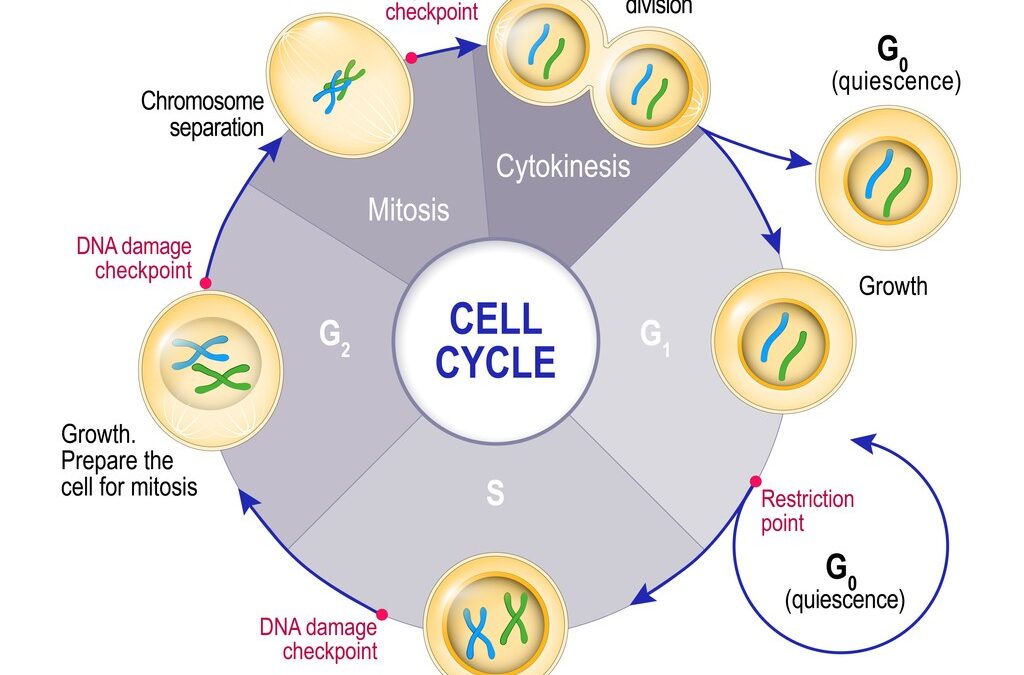
Aging is a natural physiological phenomenon wherein cells undergoing repeated division cycles, which ultimately shorten their telomere length beyond the Hayflick limit. Such aged cells may undergo self-degradation or, in some instances, adversely affect tissues of various organs, leading to open pathways for diseases to develop.


Epithalon Mechanism of Action

Epithalon, researchers suggest, increases the production of telomerase, a natural enzyme that aids in the reproduction of telomeres—protective components of DNA that prevent the loss of genetic information. Researchers speculate that this results in the prevention of chromosome shortening, which is closely linked to age-related diseases. By activating telomerase, cells may potentially exceed their predetermined time limit and reverse the mechanism of cell aging, as cells with longer telomere strands are believed to provide better function and replication.


Cell Aging and Epithalon

The cell life extension and anti-aging potential of the synthetic tetrapeptide Epithalon is considered to be strong, according to data from various experimental studies. Researchers suggest that Epithalon may achieve this by elongating telomeres and activating telomerase, supporting data that it may prolong the life of cells. Additionally, Epithalon is speculated to stimulate the endogenous secretion of melatonin, an antioxidant that may decrease age-related alterations in the immune and neuroendocrine systems, potentially reducing the incidence of chronic diseases and infections.


Disclaimer: The products mentioned are not intended for human or animal consumption. Research chemicals are intended solely for laboratory experimentation and/or in-vitro testing.  Bodily introduction of any sort is strictly prohibited by law.  All purchases are limited to licensed researchers and/or qualified professionals. All information shared in this article is for educational purposes only.

Differences of BPC-157 and TB-500 in Tissue Repair

Differences of BPC-157 and TB-500 in Tissue Repair

Peptides are short stretches of amino acids and can be either sourced from naturally prevalent proteins or synthesized. The amino acid sequences are chosen in a way that they may retain the biological function of their full-length protein counterparts and mimic their biological functions. They are said to play diverse roles from hormones, catalysts, and stimulants. The speculated role of peptides in various biological processes—whether initiating a reaction, speeding processes up, stopping the action of catalytic enzymes, or stimulating the release of hormones—varies and appears to be highly dependent on their mechanisms of action.

In this article, we will compare BPC-157 and TB-500, two peptides that have been widely researched for their potential in tissue repair processes, in terms of their chemistry, mechanism of action, and physiological influence.


Chemistry of BPC-157 and TB-500

Both BPC-157 and TB-500 are synthetic in origin, implying that they may not be found in nature and might not have common sequence homology with natural peptides. BPC-157 is a pentadecapeptide of BPC or Body Protection Compound, identified and isolated from the gastric juice. BPC-157 is a partial sequence of BPC and comprises a chain of 15 amino acids. TB-500 is synthetic form of Thymosin Beta 4, a water-soluble and regenerative compound found abundantly in bio-fluids such as saliva, and cerebrospinal fluid. TB-500 is the isolated (17)LKKTETQ(23) segment of Thymosin Beta 4 and has been researched for its potential to exert similar impacts.


BPC-157 and TB-500 Mechanism of Action

Research suggests that BPC-157 may mediate upregulation of growth hormones, modulate Nitric Oxide synthesis, enhance blood vessel production, and modify collagen production as well as bone proteins. TB-500, on the other hand, is suggested to principally associate with actin, promoting cellular migration and regeneration, and blocking inflammatory chemokines and cytokines.


BPC-157

The role of BPC-157 is centered on its potential ability to increase angiogenesis or the production of blood vessels by modifying the expression of Vascular Endothelial Growth Factor 2 (VEGFR2). This increase in angiogenesis is believed to trigger a cascade of different effects from faster wound healing, improved tendon and bone repair, and tissue regeneration in research subjects. Another suggested action of BPC-157 is its unique potential to enhance the tendon repair process and reduce the recovery time needed for damaged tendons to return to their original strength. BPC-157 is also suggested to show a cytoprotective effect, especially in the gastric mucosa, apart from the liver, pancreas, heart, and brain neurons.


TB-500

The research in TB-500 is based on its potential to bind with actin and improve tissue regeneration, tissue formation, and aid wound healing. It also appears to improve cell migration, re-epithelialization, and angiogenesis that are claimed to be crucial in tissue repair and wound healing. The peptide is also suggested to exhibit an anti-inflammatory effect by suppressing the release of chemokines and cytokines responsible for inflammation. It is currently being studied for its potential cardioprotective and neuroprotective characteristics. In-vitro tests in animal models also suggest the ability of TB-500 to inhibit premature apoptosis or cell death in the heart and the hippocampal region of the brain.


Conclusion of BPC-157 and TB-500

Based on theoretical deductions and scientific findings, both the peptides are speculated to exert action in wound healing and tissue formation. They are suggested to showcase angiogenesis and cytoprotective influence, although they may affect different factors in the process. The difference appears to be that BPC-157 influences tendon and bone repair while TB-500 doesn’t. This suggests it may potentially be more impactful in injuries that damage tendons, ligaments, and bones. It is also claimed to have greater neurological impact compared to the latter. TB-500, on the other hand, is suggested to exhibit anti-inflammatory, cardioprotective, and neuroprotective roles that BPC-157 may lack.


Disclaimer: The products mentioned are not intended for human or animal consumption. Research chemicals are intended solely for laboratory experimentation and/or in-vitro testing.  Bodily introduction of any sort is strictly prohibited by law.  All purchases are limited to licensed researchers and/or qualified professionals. All information shared in this article is for educational purposes only.
How Do Delta Sleep Inducing Peptides Work?

How Do Delta Sleep Inducing Peptides Work?

Delta Sleep-Inducing Peptide (DSIP) is an endogenous neuropeptide that was first isolated from rabbits by Monnier and associates in 1964. It is proposed to play a role in inducing delta EEG sleep and is believed to have a diverse influence on the central nervous system. Researchers suggest that it may regulate circadian rhythm, sleeping patterns, and the endocrine system while potentially alleviating hypothermia. Additionally, DSIP is speculated to protect against oxidative stress and reduce the body’s response to acute stress.

Findings from research suggest that substance P, beta-endorphin, and cortisol levels in the hypothalamus and blood plasma may show alterations under DSIP influence. It is proposed to induce changes in other peptides that mediate the acute and long-term stress coping effect. Furthermore, DSIP is believed to regulate opioid-peptidergic systems.


Delta Sleep-Inducing Peptide and Sleep, Circadian Rhythm

The peptide is said to exist in both free and bound forms in the hypothalamus, limbic system, and pituitary gland. It is believed to trigger hypothalamic neural circuitry, increasing LH during sleep, and may help in cases of chronic insomnia, according to speculative research.

A study of research models of chronic insomnia was completed to determine the intermediate effect of the peptide on sleep and daytime performance. Delta Sleep-Inducing Peptide was delivered under placebo-controlled, double-blind conditions for seven successive nights.
Polysomnograms were captured for placebo baseline, beginning and end of Delta sleep-inducing peptide exposure, and one placebo post-exposure at night. The daytime psychological state and mental performance were extensively analyzed before and after 6 Delta sleep-inducing peptide influence. Each model researched IV Delta sleep-inducing peptide, and the findings reported a greater regulation in sleep cycles.

The speculated findings also propose that DSIP may mitigate instances of narcolepsy by reducing the number of sleep attacks during the day and enhancing REM sleep. DSIP was delivered to research models of narcolepsy, with results observed to encompass decreased sleep attack frequency and improved activity, alertness, and performance during the daytime. The peptide was suggested to reduce the sleep period with the enhancement of REM sleep, accentuating circadian and ultradian rhythms.


Delta Sleep-Inducing Peptide and Opioid Withdrawal

It is speculated that DSIP might help deal with opioid withdrawal due to its suggested agonistic activity at opioid receptors. Animal study findings have suggested that DSIP, when delivered directly into the bulbo-mesencephalon-thalamic recruiting system, may trigger slow-wave sleep, and this effect may be reversed by naloxone.


Pain Perception and Depressive Behavior

DSIP is also being evaluated for potential in chronic pain and/or depression research. DSIP has been suggested to exert significant reductions in pain levels and depressive states when introduced. Due to its suggested modulating effect on endogenous opioid-peptidergic systems and its impact on circadian rhythms and cortisol levels, a study was conducted on research models of migraine episodes, vasomotor headaches, chronic tinnitus, psychogenic panic attacks, and depressive states. Delta Sleep-Inducing Peptide was suggested to remarkably lower pain levels in the majority of research models and indicated a significant decrease in depressive states following exposure.


Disclaimer: The products mentioned are not intended for human or animal consumption. Research chemicals are intended solely for laboratory experimentation and/or in-vitro testing.  Bodily introduction of any sort is strictly prohibited by law.  All purchases are limited to licensed researchers and/or qualified professionals. All information shared in this article is for educational purposes only.
What are Copper Peptides (GHK-Cu), and How Do They Work?

What are Copper Peptides (GHK-Cu), and How Do They Work?

GHK-Cu peptide is a naturally prevalent copper complex first identified in plasma, and is considered to be present in fluids such as saliva and urine. Copper peptides, small protein fragments, are believed to have a high affinity for copper ions, which are critical to normal body function. GHK-Cu peptide is thought to play essential roles, including wound healing, immune cell attraction, anti-inflammatory effects, stimulation of collagen and glycosaminoglycan synthesis in skin fibroblasts, and promotion of angiogenesis.

Speculations suggest that the peptide is secreted in response to tissue injury, aiding in protecting tissues from inflammatory damage and promoting tissue remodeling and regeneration post-injury. It is theorized to play a significant role in signaling tissue remodeling, specifically in removing damaged or scarred tissue. However there may be a reduction in its concentration, speculated to contribute to increased inflammation, cancerous activity, and tissue destruction.


Copper Peptides Peptide Mechanism of Action

GHK-Cu peptide may influence various pathways through its peptide sequence and copper. It is suggested to attract mast cells and macrophages to the injury site, releasing proteins that stimulate tissue repair. The peptide may primarily increase collagen, elastin, proteoglycans, glycosaminoglycans, and decorin in fibroblasts. It may stimulate metalloproteases and protease inhibitors to remove damaged tissue proteins and decrease the secretion of TGF-beta from fibroblasts, potentially reducing scar formation.

The speculated mechanism proposes that GHK-Cu peptide promotes collagen production by chondrocytes, leading to bone growth and formation. Additionally, it may provide copper for angiogenesis in tissues. The peptide is thought to improve the differentiation and proliferation of axons within neurons, block ferritin channels, and release oxidative iron after tissue injury, potentially blocking iron-catalyzed lipid peroxidation. Thus, copper-peptide-induced tissue repair is theorized to work for various tissues, including skin, hair follicles, stomach lining, intestinal lining, bone tissue, hooves, etc.


GHK-Cu Research Studies

Scientific work suggests that GHK-Cu peptide may affect various aspects, including infection control, wound healing, follicle growth restoration, anti-cancer cell proliferation, and neurological impact.

Wound Healing and Infection Control: Speculative findings propose that the peptide may cause better wound contraction, faster granular tissue development, and improved angiogenesis. Systemic exposure to the peptide might promote healing, and its introduction into the muscle could potentially help repair distant sites.

Anti-Inflammatory Response: Research suggests that GHK-Cu peptide might interact with TNF-alpha and pro-inflammatory cytokines, such as IL-6, in fibroblasts, leading to a significant reduction in inflammation. This may make the peptide suitable for systemic exposure in research on inflammatory skin cell conditions, such as psoriasis.

Follicle Growth Restoration: The peptide has been suggested to have efficacy comparable to other established research compounds in the restoration of follicle growth.

Cognitive Function: The peptide is speculated to promote cognitive function by increasing the migration of hematogenous cells into collagen tubes, producing nerve growth factors, increasing the expression of integrins, and enhancing the regeneration rate of myelinated nerve fibers. A gene expression study suggests that the peptide may induce changes in the expression of fundamental proteins, impacting various biochemical pathways in organs and tissues, including the nervous system.

Anti-Cancer: GHK-Cu has been suggested by researchers to be relevant in research studies of metastatic cancer. It is speculated that higher tissue copper levels may keep cells younger, and GHK-Cu may assist in mediating this effect.

Lungs: Research suggests that the peptide may protect the lungs from acute injury and fibrosis, mitigating damage due to reactive oxygen species (ROS) and inflammatory cytokines. Exposure to GHK-Cu peptide is theorized to reduce inflammatory cell infiltration and interstitial thickness in research models of pulmonary fibrosis, exhibiting potential in research areas of various lung diseases.


Disclaimer: The products mentioned are not intended for human or animal consumption. Research chemicals are intended solely for laboratory experimentation and/or in-vitro testing.  Bodily introduction of any sort is strictly prohibited by law.  All purchases are limited to licensed researchers and/or qualified professionals. All information shared in this article is for educational purposes only.
What is a Peptide?

What is a Peptide?

A peptide is a chemical compound consisting of two or more amino acids connected via peptide bonds. A peptide bond is a covalent bond. It is formed when two amino acids’ carboxyl group or C-terminus react with the amino group or N-terminus. Thus a condensation molecule of water is released during this reaction. The resulting bond is the CO-NH b.

The word peptide comes from the Greek word meaning “to digest.” Peptides are an important part of nature and biochemistry, and thousands of peptides occur naturally within the internal systems of organisms. With technological advances, new peptides are also being discovered and synthesized regularly in a laboratory settings.


The Formation Of Peptides

Peptides are formed both via natural processes within the organism and via synthetic processes inside the laboratory. Some peptides are manufactured organically, like ribosomal and nonribosomal peptides. In a laboratory, modern peptide synthesis processes may make a virtually infinite number of peptides using synthesis techniques like solid-phase or liquid-phase peptide synthesis. Solid-phase peptide synthesis is the standard process today, while liquid-phase peptide synthesis has some advantages. Vincent du Vigneaud synthesized the first Oxytocin, which is a polypeptide that was synthesized in 1953.


Classes

Peptides are divided into several classes. These classes vary based on how the peptides are produced. For example, the translation of mRNA produces ribosomal peptides. Ribosomal peptides appear to work as hormones and signaling molecules in organisms. These can include calcitonin peptides, vasoactive intestinal peptides, pancreatic peptides, and tachykinin peptide opioid peptides. Some organisms produce antibiotics like microcins, which are classified as ribosomal peptides. Ribosomal peptides mostly undergo the process of proteolysis, the breakdown of proteins into smaller peptides or amino acids to reach the mature form.

On the contrary, peptide-specific enzymes produce nonribosomal peptides. The ribosome does not produce them. Nonribosomal peptides are mainly cyclic rather than linear, although linear nonribosomal peptides may occur. Nonribosomal peptides may develop as extremely intricate cyclic structures. Nonribosomal peptides appear in plants, fungi, and single-celled organisms. Glutathione, suggested to be a significant part of antioxidant defense mechanisms in aerobic organisms, is the most common nonribosomal peptide.

Milk proteins may also form peptides in organisms. They may be produced by an enzymatic breakdown by digestive enzymes or by the proteinases formed by lactobacilli during the fermentation of milk. In addition, peptones are peptides from animal milk or meat that have been digested through proteolytic digestion. Peptones are used in the laboratory as nutrients for growing fungi and bacteria.

Moreover, fragments are commonly found as the products of enzymatic degradation in a laboratory with a control sample. However, peptide fragments can also occur naturally due to degradation by natural effects.


Terminologies to Know When Working with Peptides:

Amino Acids – Peptides consist of amino acids. An amino acid is any molecule that consists of both amine and carboxyl functional groups.

Cyclic Peptide – A peptide in which the amino acid sequence forms a ring structure instead of a straight chain. Examples of cyclic peptides are Melanotan-2 and Bremelanotide (PT-141).

Peptide Sequence – Is simply the order in which peptide bonds connect amino acid residues in the peptide.

Peptide Bond – A covalent bond formed between two amino acids when a carboxyl group of one amino acid reacts with the amino group of another amino-acid. This reaction is a condensation reaction, a reaction in which a molecule of water is released.

Peptide Mapping – Is a process that can be used to validate or discover the amino acid sequence of specific peptides or proteins.

Peptide Mimetics – Is a molecule that biologically mimics active ligands of hormones, enzyme substrates, cytokines, viruses, or other bio-molecules. Peptide mimetics can be a synthetically modified or natural peptide, or other molecules that perform the required function.

Peptide Fingerprint – It is a chromatographic pattern of the peptide. A peptide fingerprint is produced by partially hydrolyzing the peptide, which breaks up the peptide into fragments, and these are then undergone the 2-D mapping process.

Peptide Library – It consists of a large number of peptides that have a systematic combination of amino acids. Solid-phase peptide synthesis is the most frequent peptide synthesis technique that is used to prepare peptide libraries


Disclaimer: The products mentioned are not intended for human or animal consumption. Research chemicals are intended solely for laboratory experimentation and/or in-vitro testing.  Bodily introduction of any sort is strictly prohibited by law.  All purchases are limited to licensed researchers and/or qualified professionals. All information shared in this article is for educational purposes only.