Syn-AKE and Cell Regeneration

Syn-AKE and Cell Regeneration

What Is Syn-AKE Peptide?

The Syn-AKE peptide is a synthetic analog of the protein Waglerin-1. It is a 21 amino acid protein isolated from tropidolaemus wagleri. However, Syn-AKE includes only three of the amino acids which are present in the original endogenous sequence. It represents the perceived active portion of the protein and appears to produce similar action as that of the original Waglerin-1, but researchers assert that it appears to act in a controlled way.


What Are Syn-peptides?

Syn-peptides are synthetically developed peptides similar in structure to a naturally occurring peptide. They typically appear to assume the action of the naturally occurring form. Peptides are short proteins which research posits may indeed be short enough to cross the skin barrier in certain organisms.


How Does Syn-AKE Peptide Work?

Syn-AKE peptide is designed to mimic paralytic action. The Syn-AKE peptide was developed with close structural similarities to the Waglerin-1 peptide. The Waglerin-1 peptide was proposed to induce paralysis in laboratory studies, and the synthetic peptide may potentially produce a similar effect via its chemically similar structure. Researchers believe the molecule may small enough to penetrate the skin barrier. However, only tiny quantities of the molecule has been hypothesized to be able to penetrate muscle cells beneath the skin structure.

Waglerin-1 has been proposed by various researchers to induce sodium uptake by muscle cells by interacting with the mnAchR receptor. Preventing sodium uptake may block the transmission of nerve impulses to the muscles, and the muscles remain relaxed.


Disclaimer: The products mentioned are not intended for human or animal consumption. Research chemicals are intended solely for laboratory experimentation and/or in-vitro testing.  Bodily introduction of any sort is strictly prohibited by law.  All purchases are limited to licensed researchers and/or qualified professionals. All information shared in this article is for educational purposes only.

Semaglutide Peptide: Research into Gastrointestinal Function & Neurology

Semaglutide Peptide: Research into Gastrointestinal Function & Neurology

What is Semaglutide Peptide?

Semaglutide, or the Glucagon-like Peptide (GLP-1), is a naturally-occurring peptide hormone. It is a very short peptide of about 30 or 31 peptides only. The natural function of the endogenous peptide is proposed to be the regulation of blood glucose levels at an average value by interfering with natural insulin production and secretion. It appears, peculiarly, to mediate this insulin-altering function. Semaglutide peptide has been suggested by researchers to protect the beta cell insulin stores in the pancreas by upregulating the insulin gene transcription.

Apart from this main hypothesis of Semaglutide peptide, it also has neurotrophic potential on the brain and the central nervous system, which researchers are exploring in more depth. Semaglutide peptide appears to play a potentially significant role in hunger and appetite regulation in the gastrointestinal tract (GIT), potentially inducing the delay of gastric emptying and reduction of intestinal motility.

Recent studies on the Semaglutide peptide have suggested its potential action may be as far reaching as in liver, kidneys, lungs, muscles, bones, fat, and the heart function. Even though the Semaglutide peptide has mainly been investigated in the context of diabetes research, newer studies suggest its potential in cardiovascular and neurodegenerative disease research as well.

Even though this development requires additional research, some studies suggest potential in the context of Alzheimer’s disease research by slowing down the accumulation of amyloid-beta plaques in the brain, which is considered to be the primary pathology of the disease. Research conducted on the mechanism of action of the Semaglutide peptide suggested two significant mechanisms by which it appears to mediate most of its potential. These two mechanisms are described under separate headings below:


The “Incretin Effect

One of the essential hypothetical modes of action of Semaglutide- according to research conducted by Dr. Holst- is the “incretin effect.” Incretins are a group of metabolic hormones secreted by the gastrointestinal tract. The incretins are involved in decreasing the circulating blood sugar levels. Apart from gastrointestinal peptide (GIP), researchers suggest that Semaglutide or GLP-1 may potentially be the more important regulator of these hormones in the GIT.

The Semaglutide or GLP-1 receptors have been posited to exist on pancreatic beta cells, directly stimulating insulin release from the pancreas. This increased insulin secretion has been linked with other metabolic actions like increased amino acid uptake by muscle cells and increased protein synthesis and breakdown.

Studies conducted on murine models suggested that exposure to Semaglutide peptide appeared to reduce hunger levels and food intake in mice, which resulted in weight loss. This weight loss was also linked to reduced cardiovascular disease risk and decreased HbA1C levels.


Beta Cell Protection

The pancreatic beta cells are the central insulin-secreting cells. Therefore, their destruction, downregulation, or apoptosis due to any reason may possibly be a pathology of diabetes. Some compelling research on diabetic mice models has suggested that Semaglutide peptide may act upon the pancreatic beta cells in two ways. Firstly, GLP-1 may possibly stimulate the growth and proliferation of new beta cells from their progenitors in the pancreatic duct epithelium. Secondly, GLP-1 may potentially inhibit the apoptosis of these cells by inhibiting the inflammatory cytokines involved in the process. The net effect of these two actions may tip the balance towards a more significant number of pancreatic beta cells, potentially allowing them to protect the pancreas against insults that harm beta cells.


Semaglutide Peptide and The Brain

Speculative research into the peptide suggests its potentially in directing the progress of neurodegenerative diseases, specifically Alzheimer’s. It is hypothesized that the GLP-1 receptors in the brain may reduce the deposition of beta-amyloid plaques, which is the primary pathology involved in Alzheimer’s disease. Studies on rats suggested that the exposure to Semaglutide peptide appeared to improve associative and spatial learning in mice with genetic defects.


Disclaimer: The products mentioned are not intended for human or animal consumption. Research chemicals are intended solely for laboratory experimentation and/or in-vitro testing.  Bodily introduction of any sort is strictly prohibited by law.  All purchases are limited to licensed researchers and/or qualified professionals. All information shared in this article is for educational purposes only.

Overview of AICAR Peptide Research

Overview of AICAR Peptide Research

AICAR, an acronym for 5-aminoimidazole-4-carboxamide ribonucleoside, poses as a short-sequence peptide. Researchers suggest that it functions as an AMP kinase (AMPK) activator, playing a potential role in metabolic pathways and energy homeostasis. The AICAR peptide may be involved in the regulation of insulin receptors and the control of muscle cell responses to insulin.


AICAR’s Involvement in Insulin Resistance

Research study claims suggest that inflammation in adipose tissue might lead to alterations in insulin sensitivity. Research speculates that mitigating this inflammation may hypothetically enhance glucose metabolism and homeostasis without notable alterations in weight. AICAR, through unspecified pathways related to SIRT1 and macrophages, is speculated to have a role in reducing inflammation in adipose tissue. In research involving undisclosed diabetic and control mice, AICAR has been linked to a reduction in inflammatory responses by activating AMP kinase. This activation may potentially improve insulin sensitivity, energy homeostasis, lipid metabolism, and inflammatory markers.


Insights into Anti-Cancer Research and AICAR

AMPK’s conjectured impact on tumor growth and spread is presumed to be variable in different contexts. Supposedly, in certain instances, it might slow down tumor invasion, while in others, it might accentuate tumor growth. Some research implies that extended activation of the enzyme might hypothetically lead to cancer cell death by slowing cancer cell metabolism, rendering them more susceptible to environmental influences. Scientists are exploring the potential of AICAR peptides alongside other anticancer compounds to evaluate possible effectiveness against cancer cell proliferation.


AICAR Peptide and Anti-Inflammatory Action

The purported anti-inflammatory potential of AMPK activators have been the subject of exploration. AICAR, sharing potential metformin-like actions in various inflammatory conditions, is speculated to hold promise in autoimmune and inflammatory disorders. Studies in mice suggest that AICAR might decrease inflammation in colitis models. This anti-inflammatory potential may be attributed to its role as a central inhibitor of immune responses, potentially reducing nuclear factor kappa B (NF-κB) activation in macrophages and certain cytokines.


AICAR Peptide and the Heart

Inflammation of cardiovascular tissue is presumed to be the primary pathology in numerous heart diseases, including atherosclerosis. Inflammation and vascular smooth muscle proliferation may be critical factors in the failure of stent placement and other cardiovascular conditions. Therefore, controlling vascular inflammation may hypothetically reduce both short-term and long-term complications of stent placement without resorting to alternative compounds that might increase bleeding risk.

The main hypothesis that researchers hold for the AICAR peptide is that via AMPK activation, the peptide may potentially suppress specific immune responses assumed to lead to atherosclerosis. In this scenario, AICAR might mitigate the supposed risk of developing atherosclerotic plaques resulting from macrophage proliferation, hypothetically reducing the prevalence of heart diseases.


Disclaimer: The products mentioned are not intended for human or animal consumption. Research chemicals are intended solely for laboratory experimentation and/or in-vitro testing.  Bodily introduction of any sort is strictly prohibited by law.  All purchases are limited to licensed researchers and/or qualified professionals. All information shared in this article is for educational purposes only.

PTD-DBM Research in Hair Follicle Regeneration

PTD-DBM Research in Hair Follicle Regeneration

PTD-DBM peptide, or Protein Transduction Domain-fused Dishevelled Binding Motif, is the latest peptide in a series of potentially groundbreaking synthetically developed amino acid chains. It is an artificial peptide that researchers suggest might significantly influence hair follicle growth.


PTD-DBM Peptide Discovery

In South Korea, researchers conducted studies on a condition referred to as androgenetic alopecia. Research into this condition reportedly prompted Professor Choi Kang-yeol of Yonsei University and a team of researchers to explore a protein associated with follicle loss in androgenetic alopecia, namely CXXC5. The protein’s identification led to the discovery of the PTD-DBM peptide, which researchers suggest might interact with this protein to potentially alter the mechanism of follicle loss in the disease. It is also speculated to indirectly promote hair follicle growth in lab animals exposed to the peptide.


Mechanism Of Action

PTD-DBM peptide purportedly interferes with an endogenous protein, CXXC5, believed to be involved in the mechanism of hair loss. CXXC-type zinc finger protein 5 (CXXC5) is described as a negative regulator of the Wnt/β-catenin pathway, a key pathway in wound healing and hair regeneration. Allegedly, the protein CXXC5 binds to the Dishevelled protein, impeding the growth of hair follicles and leading to hair loss. This is the point at which research into the PTD-DBM peptide may become relevant, as it is suggested to activate the Wnt/β-catenin pathway by interfering with the binding of CXXC5 with the Dishevelled (Dvl) protein. The claim is that PTD-DBM may promote the growth of new hair follicles and potentially inhibit the loss of existing follicles. Moreover, there are speculative assertions about its ability to induce wound repair through wound-induced hair neogenesis (WIHN).


PTD-DBM Peptide In Wound-Healing

The Wnt/β-catenin pathway is considered common to cutaneous wound healing, dermal fibrosis, and follicle regrowth. Researchers suggest that CXXC-type zinc finger protein 5 (CXXC5) may serve as a negative feedback regulator of the Wnt/β-catenin pathway by interacting with the Dishevelled (Dvl) protein. The speculation researchers have made is that the PTD-DBM peptide may act as a common link between the protein CXXC5 and the Wnt/β-catenin pathway. Experiments on laboratory animals imply that inhibiting the CXXC5 protein might lead to accelerated cutaneous wound healing and enhanced keratin 14 and collagen synthesis. The assertion is that the PTD-DBM peptide links with the protein CXXC5, interfering with the regulation of the Wnt/β-catenin pathway and bringing about the wound-healing potential of the peptide.


Disclaimer: The products mentioned are not intended for human or animal consumption. Research chemicals are intended solely for laboratory experimentation and/or in-vitro testing.  Bodily introduction of any sort is strictly prohibited by law.  All purchases are limited to licensed researchers and/or qualified professionals. All information shared in this article is for educational purposes only.
Anti-Inflammation Studies and KPV Peptide

Anti-Inflammation Studies and KPV Peptide

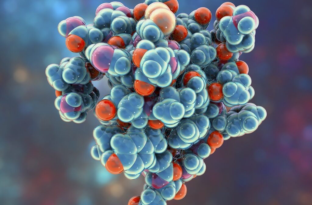
KPV is considered to be an immunomodulating peptide, naturally occurring as part of the hormone alpha-MSH. This C-terminal peptide derivative of alpha-MSH has been explored for its photoprotective characteristics, with claims that it may protect against ischemia, inflammation, and hypoxic injuries. There are also speculative assertions about its potential in feeding and energy homeostasis mechanisms. The KPV peptide, consisting of lysine-proline-valine subunits, is suggested to be a possible anti-inflammatory agent.


KPV Peptide and Inflammation

It may be suggested that KPV exhibits a pronounced anti-inflammatory effect, particularly in the small intestines, where it has been under investigation for its potential relevance in inflammatory bowel disease (IBD) research. Beyond the small intestine, researchers suggest that KPV peptide may exhibit potent anti-inflammatory effects in the central nervous system, vascular system, gastrointestinal tract, and lungs.

Research suggests that KPV may produce potential anti-inflammatory action through various mechanisms. It is speculated that KPV may potentially inactivate inflammatory pathways within cells by directly interacting with inflammatory signaling molecules that mediate the inflammatory response. It may enter the nucleus, hypothetically inactivating and halting inflammatory processes.

Speculative research proposes that this mechanism may be how it exerts a potential anti-inflammatory effect in the gut. There are suggestions that KPV might also halt pro-inflammatory mechanisms in intestinal epithelial cells and immune cells. Moreover, KPV may interact directly with immune cells, reducing inflammation. Research findings report that KPV significantly reduced inflammation in colitis, allegedly inhibiting the synthesis and secretion of pro-inflammatory cytokines, which are considered to assist in cases of IBD through inhibited immune responses.


KPV Peptide and Anti-microbial Action

Preliminary findings from research suggest that, in addition to potential anti-inflammatory effects, KPV peptide may exhibit certain antimicrobial characteristics as well. The antimicrobial activity may be targeted against two specific microbes, namely Staphylococcus Aureus and Candida Albicans. Studies suggest that KPV may inhibit the growth of S. aureus colonies.


Wound Healing and Skin Cells

Research in progress suggests that KPV peptide may potentially accelerate wound healing through speculative reductions in infection (antimicrobial) and the potential combatting of inflammation (anti-inflammatory). Research is ongoing.


Disclaimer: The products mentioned are not intended for human or animal consumption. Research chemicals are intended solely for laboratory experimentation and/or in-vitro testing.  Bodily introduction of any sort is strictly prohibited by law.  All purchases are limited to licensed researchers and/or qualified professionals. All information shared in this article is for educational purposes only.

Comparing CJC-1295 and CJC-1295 (DAC)

Comparing CJC-1295 and CJC-1295 (DAC)

Exploring CJC-1295: Considerations on Structure and Effects

CJC-1295 DAC, structurally similar to CJC-1295, features a small motif added to the end. This seemingly minor addition is speculated to have significant effects on how these peptides may function. The most crucial difference that researchers propose is its potential on the half-life of the peptide. This small alteration is speculated to increase the half-life of CJC-1295 DAC by an estimated 6 to 8 days.

CJC-1295, also known as ‘Modified GRF (1-29)’ or ‘Mod GRF 1-29,’ is a synthetic substitute for Growth Hormone Releasing Hormone (GHRH). Researchers propose that due to its structural resemblance to GHRH, Mod GRF 1-29 might induce a significant increase in growth hormone (GH) levels. Growth Hormone, a hormone produced by the thalamus in the brain, appears to control various physiological functions, including glucose metabolism, lipid breakdown, bone development, skin structure, and blood pressure regulation. Growth hormone production is considered to level off and decline naturally. Due to its speculative ability to mimic GHRH, Mod GRF 1-29 may potentially act on the growth hormone-releasing receptors of the brain, signaling the brain to continue to produce growth hormones.


CJC-1295 with DAC:

CJC-1295 with DAC, as researchers suggest, shares a structural similarity with CJC-1295, differing only in the addition of ‘Drug Affinity Complex’ (DAC) at the peptide’s end. DAC, composed of a few nitrogen and oxygen atoms, is speculated to significantly influence the biological processing of CJC-1295 DAC compared to CJC-1295. The primary distinction between the two molecules revolves around their effects on half-life. Mod GRF 1-29, with a structure akin to GHRH, is reputed to exhibit a short half-life, lasting only around 30 minutes. In contrast, researchers propose that CJC-1295 DAC, featuring the DAC end, may potentially bind to albumin proteins. This purported small addition to Mod GRF 1-29 is speculated to cause a substantial increase in its half-life, estimated to be approximately 6 to 8 days compared to the CJC-1295 peptide.


Disclaimer: The products mentioned are not intended for human or animal consumption. Research chemicals are intended solely for laboratory experimentation and/or in-vitro testing.  Bodily introduction of any sort is strictly prohibited by law.  All purchases are limited to licensed researchers and/or qualified professionals. All information shared in this article is for educational purposes only.